Overview of Musculoskeletal Anatomy/Lesson 1
Appearance
Overview of Musculoskeletal Anatomy - Lesson 1
Bone and muscles of the head and neck
| Overview of Musculoskeletal Anatomy | |
|---|---|
| Lesson: | Lesson 1 |
| Level: | Undergraduate |
| Suggested Prerequisites: | Introduction to Regional Anatomy |
| Time Investment: | 30mins |
| Assessment Methods: | Quiz |
| Portal: | Science |
| School: | Biology/Medicine |
| Division: | Anatomy |
| Department: | Regional Anatomy |
| Lesson Coordinator: | Doctorbee (talk) |
Skull
[edit | edit source]The skull, also known as the cranium, contains 22 bones. There are two sets of bones in the skull: eight cranial and fourteen facial bones.
Cranial Bones
[edit | edit source]There are eight cranial bones. They form the cranial cavity, which functions to hold and protect the brain.
- Frontal (1)
- Parietal (2)
- Temporal (2)
- Occipetal (2)
- Sphenoid (1)
- Ethmoid (1)
- Inferior nasal conchae (2)
- Vomer (1)
Frontal Bone
[edit | edit source]-
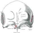
Lateral and posterior views of the frontal bone
There are two portions to the frontal bone: horizontal portion and vertical portion.
- Horizontal portion is also known as pars orbitalis and consists of two thin triangular plates, the orbital plates and ethmoidal notch, which form the vaults of the orbital and nasal cavities respectively. There are two surfaces to the pars orbitalis.
- The inferior surface is smooth and has a depression called lacrimal fossa on the lateral part. The lacrimal fossa houses the lacrimal gland, which secrete tears. There is a depression near the nasal part, and it is called the fovea trochelearis, where the obliquus oculi superior muscle attaches. This muscle is one of the muscles involved in the rolling of the eyes.
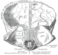
- The superior surface has depressions and contain the convolutions of the frontal lobes of the brain and the anterior and posterior ethmoidal arteries. The superior surface also contains ethmoidal notch, which separates two orbital plates.
-
Anterior view of the frontal bone
-
Posterior view of the frontal bone
- Vertical portion is also known as squama frontalis. It corresponds with the forehead region. There are two surfaces to the squama frontalis as well: external surface and internal surface.
- External surface of the squama frontalis
| Completion status: Ready for testing by learners and teachers. Please begin! |


