Motivation and emotion/Book/2017/Neurotransmitters and emotion
What is the effect of neurotransmitters on emotion?
Overview
[edit | edit source]Emotions are key providers for the motivational establishment of goal-directed behaviour, and are exceedingly entangled with our cognitive, perceptual processes. Partially due to limitations in neuroimaging techniques, the underlying neural mechanisms of emotions at present have not been able to fully explain how neural activity links to specific emotional states. Consequently, the content of neurobiology can account for emotion in a broad framework within the human brain, which describes the emotional experience in terms of a physiological, and mental representation. This chapter looks at the role of chemical messengers (neurotransmitters) within the brain, and their role with certain brain areas, in the formation of emotions.
Neurotransmitters
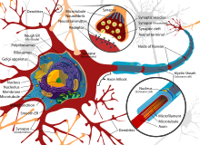
[edit | edit source]Fundamentally, neurotransmitters are certain types of hormones that comprise of amino acids located in the brain, which convey information from one neuron to another neuron. They have the ability to control our capability to experience pain and pleasure, our movement, and our emotional response to stimuli. The most common neurotransmitters that play a role in emotion are dopamine, gamma-aminobutyric acid (GABA), serotonin, and noradrenaline. Neurotransmitters can generally be categorised into two categories; inhibitory or excitatory, with some being able to serve both functions depending upon the type of receptor at the postsynaptic neuron.
How neurotransmitters work
[edit | edit source]
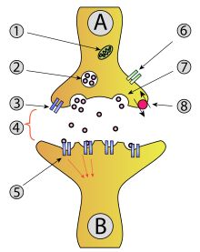
To gain a better understanding of how neurotransmitters affect our emotions, a brief explanation of how neurotransmission works in our bodies is provided. To begin with, an electrical signal (nerve impulse) travels along a neural pathway until it reaches the end of the pathway. Upon reaching the end of the pathway, a conversion of the electrical signal into a chemical signal (neurotransmitter) takes places in the synapse (space between neurons). This signal then will then cross the synapse between the neurons, where it will once again be converted to an electrical signal. An action potential triggers the release of theses neurotransmitters from the presynaptic nerve terminal, through a process of exocytosis. Neurotransmitters are essentially bundled in vesicles in the presynaptic neuron (first neuron), where upon release enter the synapse, where they then will bind to receptors on the postsynaptic neuron (second neuron). It is worthwhile to note that the release of neurotransmitters can also occur through graded electrical potentials, and a low level can be released in the absence of an action potential.
A more detail link is provided to further explain the process.
Inhibitory neurotransmitters
[edit | edit source]Inhibitory neurotransmitters reduced the probability of an excitatory signal being sent, and act as the human body’s nervous system off switch. The careful balance between excitation and inhabitation is critical as too much excitement can lead to insomnia, restlessness, and irritability. These inhibitory neurotransmitters play a role in the body to diminution of aggression, encourage calmness, and induce sleep, which in turn influence our emotions.

Excitatory neurotransmitters
[edit | edit source]Excitatory neurotransmitters play the opposite role to inhibitory neurotransmitters as they promote the probability of an excitatory signal being sent, and act as the human body’s nervous system on switch. They are responsible for energy, alertness, motor movement, fight or flight response, and higher order thinking.
- Dopamine
- Noradrenaline
Dopamine
[edit | edit source]Dopamine is involved in the brain’s reward system, which generates the feeling of pleasure, and acts as the brains feel good neurotransmitter. It has the capability to act as either an inhibitory or excitatory neurotransmitter; depending upon the particular site it binds to, and can alter our motivation, and emotional states. Once dopamine is produced, it can be transformed into the brain chemicals norepinephrine and epinephrine, and is associated with positive stress states such as sex, exercise, and being in love. The amino acids tyrosine and phenylalanine, along with B vitamins, can influence the production of dopamine.
As dopamine levels are increased when we partake in activities that bring us satisfaction and pleasure, too high of a level can become detrimental for our health. Disproportionate higher levels can cause us to become hyper-stimulated to our surrounding environment, cause a disrupted thought process, which are all characteristics of schizophrenia. A shortage of dopamine in the cortex mixed with an excess of dopamine in the limbic system, can possibly lead to suspicious thinking, and paranoia. This narrows and intensifies our focus, which can lead to mood swings, and emotional instability.

In the frontal lobe, dopamine regulates the incoming flow of information from other brain areas. An increase of dopamine in the frontal lobes produces feelings of pleasure and dismisses pain, whereas low levels in the frontal lobe reduce memory function. Consequently, low levels are associated with a lack of focus, cognitive problems such as memory and learning deficits, inability to feel alive, compulsions, addiction, and a loss of satisfaction in previously enjoyable activities. This can have a substantive impact upon an individuals emotional state.
Essentially, there are eight major dopaminergic pathways located in the brain, with three pathways starting from the midbrain, which can alter our motivation and emotion. Firstly, there is the mesolimbic pathway, which is associated with the reward circuit, and is involved in motivation and memory. Secondly, there is the mesocortical pathway, which through dysfunction may be the cause for disordered thinking, and hallucinations. Thirdly, the tuberoinfundibular pathway, which is accountable for behavioural aspects, executive functions, and emotional processing.
Noradrenaline
[edit | edit source]Noradrenaline is another neurotransmitter in the human body, which is regularly associated with the body’s fight or flight response to stress. Noradrenaline has the capacity to influence our physical responses, create a sense of urgency, overpowering fear, and an increase in heart rate and blood pressure (Moore, Summer, & Bloor, 1984; Passetti, Dalley, O'Connell, Everett, & Robbins, 2000). Emotionally, this neurotransmitter level in the brain is related to both depression, and anxiety, with a role seemingly appearing to uphold the balance between depression and nervousness. Almost all anxiety disorders involve an elevation of noradrenaline.

As noradrenaline is the neurotransmitter of arousal, low levels are linked with poor memory, depression, below average levels of alertness, and interest. Additionally, moderately high levels of noradrenaline produce uncomfortable feelings of arousal which strongly influence physical changes, intensify anxiety, impair concentration, and increases the startle reflex. This system begins from the locus coeruleus, lateral tegmental area located in the brainstem, in which through axonal pathways to the cortex, reaches the central nervous system(CNS).
Serotonin
[edit | edit source]Another neurotransmitter is serotonin, which was first isolated in 1933, with its role in various psychiatric disorders being discovered over the years. Serotonin is synthesized from the amino acid L-tryptophan in the neurons of the brain, and stored in vesicles. Serotonin is a critical regulator of bodily functions such as libido level, sleep, body temperature, and is often associated with depression, social anxiety, eating disorders, and obsessive-compulsive disorder (OCD).
When levels of this neurotransmitter are low, an individual would experience problems with attention, become unorganised, and lack concentration. As serotonin levels further decrease, simple everyday tasks become overwhelming due to the lack of ability to plan.

Consequently, this produces an individual to experience negative emotional states. When levels of this neurotransmitter are high, this can result in a life-threatening disorder known as Serotonin Syndrome (SS). SS produces malignant hyperthermia, over-confidence, aggression and agitation, along with serval physiological symptoms. Likewise to dopamine, substances received by the body can influence serotonin levels in the brain. Serotonergic cell bodies originate generally in the midbrain and upper pons area, with neurons projecting to the cerebral cortex, basal ganglia, and the limbic system (Kaplan, Middleton, Urban, & Midgley, 2002).
Gamma-Amino Butyric Acid (GABA)
[edit | edit source]The role of the neurotransmitter GABA is that which is inhibitory, meaning it reduces the ability of other neurotransmitters to work. Essentially, rather than encouraging communication between cells such as the three previously mention neurotransmitters, GABA dejects, and blocks communication. Fundamentally, GABA is involved in our level of excitement, which is essential in brain areas involving emotional states.

When levels of this neurotransmitter are low in the brain, an individual will be overstimulated, causing communication to become uncontrollable. Consequently, when levels are high in the brain, the individual will experience impaired reactions, excessively relaxed, and even become sedated. Additionally, when levels of the neurotransmitter in the brain are within normal stable ranges, the individual would not be anxious or excessively aroused, with suitable reactions to the surrounding environmental situation. Principally, the neurotransmitter GABA acts as the body’s communication speed controller, ensuring brain communication is operating at the correct intensity, and speed, to suit the situational environment around the individual.
|
Topic review: Part one
|
Emotions in specific brain areas
[edit | edit source]Through neurons in the CNS, the brain is able to communicate with the different systems of the body. Whilst the various areas of the CNS have specialised functions, the neurons themselves are unable to transmit specific information. Essentially, the body requires neurotransmitters in order for the messages from neurons and cells, to be transmitted, regulated, or amplified.
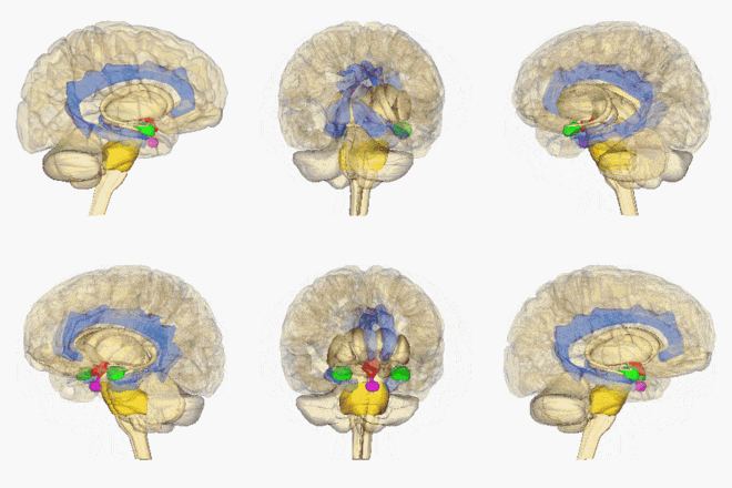
Neurotransmitters are made from different chemicals, which are released in the synapse or presynaptic nerve terminal, which play an important role in mood regulation, and emotion. The various neurotransmitters play a role within the below brain areas.
The cerebral cortex
[edit | edit source]The top layer of the brain is known as the cerebral cortex. There are large convolutions that are called fissures, which divide the brain into five lobes. They consists of the temporal lobe that is associated with hearing and through its lower levels, involved with aspects of emotional learning and memory. The occipital lobe, which is associated with vision. The parietal lobe, which is involved with body image and sensation, and the frontal lobes which are involved in movement and planned action. Through mapping techniques, the particular sensory and motor functions of the cerebral cortex have been located, with the exception of the frontal cortex, which is harder to map.
However, the frontal cortex has shown areas associated with the integration of sensory information, and the conscious registration of emotion from inner structures of the brain.
The neocortex
[edit | edit source]The neocortex is the exterior layer of the cerebral cortex, which contains association cortexes, the sensory cortex, and motor areas (Turner, 1999). LeDoux (1996), claims that higher brain functions of the cortex are critical for the production of emotional feelings. Lower level emotional structures regulate higher neural structures, which has complex ways of organising, inhibiting, and controlling our emotion.
The cingulate cortex
[edit | edit source]The cingulate cortex consists of a strip that runs from the anterior of the corpus callosum to the posterior of the callosum. The posterior of the cortex is particularly concerned in emotion, which includes transient sadness, and depression, whereas the anterior is associated with cognition. The cingulate cortex is critical in the somatosensory system, which creates feelings such as sexual excitement, reactions to recreational drugs, and arousal. As stated by Damasio (1994), the cingulate cortex role is essential in the body feeling itself, thus vital to emotion. Without the feeling from our bodies, emotion is essentially indistinguishable from thought (Carter, 1999).
Insula
[edit | edit source]The insula is located deep inside the temporal lobe, and is also an integral part of the somatosensory region for the processing of emotional feeling. When an emotional feeling is felt from the body, the signal coveys from the brain stem, to a particular nucleus of the thalamus, then to neural maps in the insula. From here, the signal is sent onto the cingulate cortex, and ventromedial prefrontal cortex.
Amygdala
[edit | edit source]
The amygdala is located at the front of the hippocampus, is known for being the brains alarm system, and has an important role in the consolidation of emotionally significant memories (Kandel, Schwartz, & Jessell, 2000). Jean-Marc, Armony, and LeDoux (2000) also claims the amygdala stores unconscious memories in a similar manner to how the hippocampus stores explicit long-term memories. Furthermore, the amygdala plays an important role in the interpretation of other people’s emotions, which coordinates both the endocrine, and autonomic systems involved in emotion. Additionally, the amygdala monitors the outside world for danger with the capability to engage in instant emotional states such as anger, and fear. As the amygdala controls autonomic responses associated with the fear network, it has been hypothesised that medications, which increase the amount of the available neurotransmitter serotonin, would essentially desensitise the fear network. When scientists artificially stimulated the amygdala in patients, they reported increased feelings of fearfulness, and anxiousness. Additionally in animal studies, scientists executed a lesion to destroy the amygdala and found this resulted in the animals becoming hyper-sexual with a calmer emotional state (Wootton, Frick, Shelton, & Silverthorn, 1997).
Hippocampus
[edit | edit source]The hippocampus facilitates in the storage, and learning of important emotional lessons. As this provides emotional conditioning, without the ability to be able to recall previous experiences, would result in an individual experiencing a limited development in emotional intelligence. Additionally, it is responsible for spatial orientation, and navigation. The hippocampus has the ability to recognise both the danger, and the contexts of a situation or object, and works closely with the amygdala. Being connected to almost the whole cortex, the hippocampus aids in memory consolidation.
The diencephalon
[edit | edit source]Between the midbrain and the cerebral hemispheres lies the diencephalon, which is composed of the thalamus and the hypothalamus. Accordingly to Turner (1999), the pituitary gland and the diencephalon monitor the emotional charges produced from sensory inputs. Furthermore, both areas produce peptides, and hormones essential to emotional response.

The thalamus acts essentially like a relay station for the periphery system, distributing, and processing all motor and sensory information. Kandel, Schwartz, and Jessell (2000) found that the thalamus acts to govern this information, whether it actually reaches awareness. The thalamus also acts as an integrating center, as it has the ability to modify incoming information.
The hypothalamus is involved in the chemical responses that comprise emotion, with input and output connections to regions in the central nervous system, responsible to emotional feeling. The hypothalamus is the center for homeostasis, which performs multiple functions such as activating the sympathetic nervous system, secretes trophic hormones which control the release of hormones from the anterior pituitary gland, and involved in the interaction with the limbic system to influence our emotions and behaviour (Wootton, Frick, Shelton, & Silverthorn, 1997).
The olfactory cortex
[edit | edit source]The olfactory cortex receives feedback from chemoreceptors located in the nose, and is located in the temporal lobe of the brain. Olfactory information is the only sensory information that is not sent through the thalamus, instead sent to the olfactory bulb in the cerebrum. Through this direct feedback from the olfactory bulb to the cerebrum, it is hypothesised that this is the reason for nostalgia, as a simple smell can trigger the reminiscence of pervious memories and associated emotions.
|
Topic review: Part two
|
Silvan Tomkins "Affect Theory"
[edit | edit source]
Silvan Tomkins developed a comprehensive theory of basic emotions, in which he classified eight sets of emotions. In the formation of the emotional sets, he labeled one word for low intensity, and the other for high intensity of the emotion (Tomkins, 1982; Tomkins & McCarter, 1964). He referred to these basic emotions from a biological standpoint (Nathanson, 1992), which consists of one neutral set; Surprise/Startle, two positive sets; Enjoyment/Joy and Interest/Excitement, and five negative sets; Anger/Rage, Fear/Terror, Contempt/Disgust, Distress/Anguish, and Shame/Humiliation. Regarding the neutral set, Edman (Edman, Friesen, & Simons, 1985) influenced Tomkins that startle was unconnected to surprise, hence the neutral set now comprises solely of Surprise.
Enjoyment/Joy and Interest/Excitement
Enjoyment/Joy is in the low-noradrenergic corner, whilst being high in both serotonin, and dopamine. Individuals in this state experience emotions that are generally relaxed and calming. Interest/Excitement has all three monoamines showing as high. Dopamine plays a significant role in exploratory activity (Mallo et al., 2008), appetite (Faure, Reynolds, Richard, & Kerridge, 2008), love (Ortigue, Bianchi-Demicheli, Patel, Frum, & Lewis, 2010), and addiction (Ross & Peselow, 2009). Noradrenaline has been found to elevate with the initiation of food (Ventura, Latagliata, Morrone, & La Mela, 2008), receiving positive food rewards (Young & Williams, 2010), and sexual excitement (Rodriguez-Manzo & Fernandez-Guasti, 1995). Regarding the serotonergic properties, this is supported by the effects of recreational drugs such as ecstasy (Bedi, Hyman, & de Wit, 2010). Both these two sets of emotions are classified according to Tomkins as being positive (Tomkins & McCarter, 1964).
Surprise
Surprised is in the low-dopaminergic, high-noradrenergic, and high-serotonergic corner, and is described as a neutral basic emotion.
Anger/Rage and Fear/Terror
These two sets of emotions are linked to reinforcement through being high in dopamine (Fadok, Darvas, Dickerson, & Palmiter, 2010). Furthermore, the rewarding effects of these two sets of emotions might explain why people engage in risky behaviour to deliver an adrenaline rush. This has been supported with the study of laboratory rats, in which the rats learnt to avoid scary stimuli (Phelps & LeDoux, 2005), and through studies on patients with Parkinson’s disease. One lot of patients showed a blunted response to aversive stimuli (Bowers et al., 2006), whilst another group of patients showed an impairment in the ability to recognise angry faces after the dopamine replacement theory was withdrawn.
These two sets of emotions are also linked to individuals who are feeling threatened or under pressure, resulting in a low serotonin level. Moreover, through studies on aggression and anger, this supports the idea of the low serotonin properties (Seo, Patrick, & Kennealy, 2008). Additionally patients with Alzheimer’s disease showing aggression have been treated with selective serotonin re-uptake inhibitor's(SSRI) (Barak, Plopski, Tadger, & Palace, 2011).
Fear/Terror has been associated with being low in noradrenaline, with it being linked to a cold fear where the heart slows down. The fight or flight reaction is linked more with the Anger/Rage basic emotion, as this is associated with higher noradrenaline levels such as an individual having a higher heart rate, being aroused, and active (McGaughy, Ross, & Eichenbaum, 2008).

Contempt/Disgust
Contempt/Disgust is linked with being high in serotonin, and low in both dopamine and noradrenaline. A core feature in disgust is food related disgust, as once the individual feels satisfied, they will experience a feeling of aversion towards food. Moreover due to tryptophan depletion, the ability to recognise disgusted faces is reduced (Aan Het Rot, Coupland, Boivin, Benkelfat, & Young, 2010). The relationship between Contempt/Disgust and Shame/Humiliation is best described as contempt for an object versus self-contempt. Furthermore, as disgust is associated with low dopamine, this links to withdrawal and repulsion. Patients with anhedonia and schizophrenia have been found to experience greater levels of disgust (Ille, Schony, Kapfhammer, & Schienle, 2010), whilst animal studies have shown that a conditioned taste aversion stimulus results in a decrease of dopamine in the nucleus accumbens (Mark, Blander, & Hoebel, 1991).
Distress/Anguish and Shame/Humiliation
In Shame/Humiliation, all three monoamines are low. Tomkins (1981) claimed that shame strikes into the deepest heart of man, with the individual suffering feelings of unworthiness, and lack of dignity. Distress/Anguish is similar to Shame/Humiliation, and believed to be the equivalent to Shame/Humiliation as serotonin and dopamine are low, however has high levels of noradrenaline. Current findings support the relationship between SSRI antidepressants on anxiety disorders (Schneider, 2006), depression and anxiety (Pollack, 2005), and depression and shame (Vikan, Hassel, Russet, Johansen, & Moen, 2010), with an individual being low in serotonin. Additionally, patients with generalised anxiety disorder, particularity social phobias, have been found to be low in dopamine (Fergus, Valentiner, McGrath, & Mencius, 2010).
Other relevant theories of emotion
[edit | edit source]There are numerous other theories that endeavour to explain the complex relationship between distinct brain regions, and the link to emotional states:

James-Lange theory of emotion
This theory of emotion proposes that physiological change is primary, followed by an emotional response as the brain reacts to the information received from the nervous system. To account for the various types of emotional experiences, it is claimed that neuro activity in the autonomic system as a results of a particular stimuli, triggers an emotional expression in the brain.
More about the James-Lange theory can be found on the link provided.
Cannon-Bard theory of Emotion (Thalamic Theory)
This is another theory of emotion, which propses that while the physiological response does play an important role in our emotions, the physiological response alone cannot entirely explain the full spectrum of an emotional experience. Emotions are not simply a result of a particular bodily state as previous thought, as the physiological response is too slow, and unable to account for the intense, rapid awareness of an emotion. As an individual learns new information about a specific experience or event, the initial visceral reaction can vanish. The previous theory, which results from a chemical perspective, simply cannot account for cognitive modulation of emotion as new information is learnt.
More about the Cannon-Bard theory can be found on the link provided.
The Schachter-Singer Theory (Two-Factor Theory)
This particular theory can be linked to the Cannon-Bard theory, as it claims that physical arousal can commonly have no intrinsic meaning, and to arrive at an emotional state we rely on information from the situation. Schacter and Singer in 1962 conducted a study where they divided the subjects into groups based on the administration of adrenaline into the test subjects veins, and tried to induce either euphoria or anger. The results showed that the subjects which knew they had received adrenaline, despite having elevated physiological responses, did not become as euphoric or angry when compared to the subjects who were unaware. This provided further support for the cognitive elevaluation of a stimulas being more important than solely a physiological change, in the emotional experience.
More about the Schachter-Singer theory can be found on the link provided.
Neurotransmitters and emotional disorders
[edit | edit source]
Depression
[edit | edit source]In regards to the neurotransmitters dopamine, serotonin, and norepinephrine, depression has been associated with the brain having an imbalance of chemicals. Currently, it is challenging to accurately measure such neurotransmitters in the brain, however antidepressant medications have been shown to impact upon the specific receptors, and neurotransmitters.
Concerning the neurotransmitter dopamine, this plays a critical role in our ability to achieve pleasure, regulates our motivation to pursue rewards, and has been associated with depression. When dopamine levels are low, generally depressed individuals will not develop the equivalent sense of pleasure when partaking in original activities, which once brought pleasure. Additionally, the neurotransmitter serotonin has also been associated with depression. Serotonin is produced by serotonergic neurons, and is involved in regulating various significant physiological functions, and mood. When the production of serotonin is decreased in the serotonergic neurons, current research suggests this can cause individuals to experience a negative emotional state, with increased feelings of suicide.
Furthermore, the neurotransmitter noradrenaline has been associated with depression. This dates back to the catecholamine hypothesis of affective disorders (Schildkraut, 1965), developed in the 1960s, as an explanation as to why individuals become depressed. Essentially this hypothesis proposes that it is a deficiency of this neurotransmitter in the brain, which is accountable for negative emotional states. Current findings have indicated that while there are lower levels of the neurotransmitter in some depressed individuals, lower levels of noradrenaline do not always cause an individual to experience a negative emotional state.

Consequently, there has been cases which have pointed at some individuals as having higher levels of noradrenaline due to hyperactivity of the noradrenergic neurons, with additional research proposing that it is the drop in serotonin levels which fundamentally causes a drop in noradrenaline levels, resulting in negative emotional states.
Though further research is required to illustrate the particular impact of the neurotransmitter glutamate has on depression, glutamate transmission mediates the emotion and cognition of brain functions (Pessoa, 2008). Through recent research by Robbins and Arnsten (2009), has concluded that monoaminergic transmission plays a critical role in the variation of emotion and cognition, current antidepressant contain either a monoamine-base component or monoamine-based mechanism, but none directly target the glutamatergic system (Sanatoria, Zaeate, Krystal, & Manji, 2008). The findings for the largest open-label study contacted by Trivedi et al., (2006) has shown that only 50-60% of depressed individuals respond to monoamine-based mechanisms with less than one third achieving remission.
Anxiety
[edit | edit source]Anxiety is characterised as a frequent negative emotional state, which results in feelings of apprehensions and uneasiness, being accompanied with behavioural and cognitive expressions. Through various branches of neuroscience, it is evident that the dysfunction in the modulation of brain circuits, which control emotion responses, can result in anxiety disorders. This can range from top-down processing where the stimuli which triggers the emotional response, originates in the prefrontal cortex, to bottom-up processing originating from the amygdala.
As serotonin has been identified for its influence on mood, sleep, and appetite, it is commonly associated with anxiety disorders (Durant, Christmas, & Nutt, 2010; Gordon & Hen 2004). Medications such as SSRI's are regularly prescribed to individuals who have a decreased level of serotonin.

Likewise to serotonin, GABA has also been associated to anxiety, as this works to quiet the brain, relax the body, and slow down neural transmission. For individuals who experience chronic anxiety, it is proposed that a GABA deficiency may be the reason for increased anxiety, as the individuals bodily functions are already in a heightened state of attentiveness, and arousal. Consequently, this may mean the individual is biological vulnerable to anxiety.
Conclusion
[edit | edit source]Whilst we have a somewhat thorough understanding of the mechanisms of the brain, emotions are extremely complex processes which still remain poorly understood. Emotions arise from a variety of different elements, including social relationships, automatic behaviours, physiological responses, environmental cues, and cognitive interpretations. Neurotransmitters are simply one mechanistic component involved in the dynamic processes which shape our emotional response, and experiences. Emotions are not single entities, and neurotransmitters do not directly cause emotions. Whilst brain chemistry and emotions are interconnected, saying that neurotransmitters cause emotion, and emotional processing affects neurotransmitters, essentially down plays the complexity of the human brain.
See Also
[edit | edit source]- Discrete emotion theory
- Lövheim cube of emotion
- Neurotransmitters and motivation (Book chapter, 2017)
- Norepinephrine and motivation (Book chapter, 2017)
- Norepinephrine and emotion (Book chapter, 2017)
References
[edit | edit source]Barak, Y., Plopski, I., Tadger, S., & Paleacu, D. (2011). Escitalopram versus risperidone for the treatment of behavioral and psychotic symptoms associated with Alzheimer's disease: A randomized double-blind pilot study. International Psychogeriatrics, 1-5.
Bedi, G., Hyman, D. H., & de Wit, I. (2010). Is ecstasy an empathogen? Effects of +/- 3, 4-methylenedioxymethamphetamine on prosocial feelings and identification of emotional states in others. Biological Psychiatry, 68, 1134-1140.
Bowers, D., Miller, K., Mikos, A., Kirsch-Darrow, L., Springer, U., & Fernandez, H. (2006). Startling facts about emotion in Parkinson's disease: Blunted reactivity to aversive stimuli. Brain, 129, 3356-3365.
Carter, R. (1999). Mapping the Mind. Berkeley: University of California Press.
Damasio, A. (1994). Descartes' Error: Emotion, Reason, and the Human Brain. New York: Avon Books.
Durant, C., Christmas, D., & Nutt, D. (2010). The pharmacology of anxiety. Current Topics in Behavorial Neirosciences, 2, 303-330.
Ekman, P., Friesen, W. V., & Simons, R. C. (1985). Is the startle reaction an emotion? Journal of Personality and Social Psychology, 49, 1416-1426.
Fadok, J. P., Darvas, M., Dickerson, T. M., & Palmiter, R. D. (2010). Long-term memory for pavlovian fear conditioning requires dopamine in the nucleus accumbens and basolateral amygdala. PLoS One, 5, 12751.
Faure, A., Reynolds, S. M., Richard, J. M., & Berridge, K. C. (2008). Mesolimbic dopamine in desire and dread: Enabling motivation to be generated by locakized glutamate disruptions in nucleus accumbens. Journal of Neuroscience, 28, 7184-7192.
Fergus, T. A., Valentiner, D. P., McGrath, P. B., & Jencius, S. (2010). Shame and guilt proneness: Relationships with anxiety disorder symptoms in a clinical sample. Journal of Anxiety Disorders, 24, 811-815.
Gordon, J. A., & Hen, R. (2004). The serotonergic system and anxiety. Neuromolecular Medicine, 5(1), 27-40.
Ille, R., Schony, M., Kapfhammer, H. P., & Schienle, A. (2010). Elevated disgust proneness in schizophrenia. Journal of Clinical Psychology, 66, 1090-1100.
Jean-Marc, F., Armony, J. L., & LeDoux, J. E. (2000). Emotional Circuits. In M. Lewis, & J. M. Haviland-Jones, The Handbook on Emotions (pp. 398-401). New York: Guilford.
Kandel, E. R., Schwartz, J., & Jessell, T. M. (2000). Principles of Neural Science. New York: McGraw-Hill.
Kaplan, A., Middleton, M. J., Urdan, T., & Midgley, C. (2002). Achievement goals and goal structures. In C. Midgley, Goals, goal structures, and patterns of adaptive learning (pp. 21-53). Mahwah: Lawrence Erlbaum.
LeDoux, J. (1996). The Emotional Brain: The Mysterious Underpinnings of Emotional Life. New York: Simon and Schuster.
Mallo, T., Alttoa, A., Koiv, K., Tonissaar, M., Eller, M., & Harro, J. (2007). Rats with persistently low or high exploratory activity: Behaviour in tests of anxiety and depression and extracellular levels of dopamine. Behavioural Brain Research, 177, 269-281.
Mark, G. P., Blander, D. S., & Hoebel, B. G. (1991). A conditioned stimulus decreases extracellular dopamine in the nucleus accumbens after the development of a learned taste aversion. Brain Research, 551, 308-310.
McGaughy, J., Ross, R. S., & Eichenbaum, H. (2008). Noradrenergic, but not cholinergic, deafferentation of prefrontal cortex impairs attentional set-shifting. Neuroscience, 153, 63-71.
Moore, N. C., Summer, K. R., & Bloor, R. N. (1984). Do patients like psychometric testing by computers. Journal of Clinical Psychology, 40(3), 875-877.
Nathanson, D. (1992). Affect, sex, and birth of the self. New York: Norton and Company.
Ortigue, S., Bianchi-Demicheli, F., Patel, N., Frum, C., & Lewis, J. W. (2010). Neuroimaging of love: fMRI meta-anakysis evidence toward new perspectives in sexual medicine. The Journal of Sexual Medicine, 7, 3541-3552.
Passetti, F., Dalley, J. W., O'Connell, M. T., Everitt, B. J., & Robbins, T. W. (2000). Increased acetylcholine release in the rat medial prefrontal cortex during performance of a visual attentional task. European Journal of Neuroscience, 12, 3051-3058.
Pessoa, L. (2008). On the relationship between emotion and cognition. Nature Reviews Neuroscience, 9, 148-158.
Phelps, E. A., & LeDoux, J. E. (2005). Contributions of the amygdala to emotion processing: From animal models to human behavior. Neuron, 48, 175-187.
Pollack, M. H. (2005). Comorbid anxiety and depression. Journal of Clinical Psychiatry, 66, 22-9.
Robbins, T. W., & Arnsten, A. F. (2009). The neuropsychopharmacology of fronto-executive function: Monoaminergic modulation. Annual Review of Neuroscience, 32, 267-287.
Rodriguez-Manzo, G., & Fernandez-Guasti, A. (1995). Partcipation of the central noradrenergic system in the reestablishment of copulatory behavior of sexually exhausted rats by yohimbine, naloxone, and 8-OH-DPAT. Brain Research Bulletin, 38, 399-404.
Ross, S., & Peselow, E. (2009). The neurobiology of addictive disorders. Clinical Neuropharmacology, 32, 269-276.
Sanacora, G., Zarate, C. A., Krystal, J. H., & Manji, H. K. (2008). Targeting the glutamatergic system to develop novel, improved therapeutics for mood disorders. Nature Reviews Drug Discovery, 7, 426-437.
Schildkraut, J. J. (1965). The catecholamine hypothesis of affective disorders: A review of supporting evidence. The American Journal of Psychiatry, 122(5), 509-522.
Schneier, F. R. (2006). Clinical practice. Social anxiety disorder. The New England Journal of Medicine, 355, 1029-1036.
Seo, D., Patrick, C. J., & Kennealy, P. J. (2008). Role of serotonin and dopamine system interactions in the neurobiology of impulsive aggression and its comorbidity with other clinical disorders. Aggression and Violent Behavior, 13, 383-395.
Tomkins, S. (1982). Affect theory. In P. Ekman, W. Friesen, & P. Ellsworth, Emotions in the human face (pp. 355-395). Cambridge: Cambridge University Press.
Tomkins, S., & McCarter, R. (1964). What and where are the primary affects? Some evidence for a theory. Perceptual and Motor Skills, 18, 119-158.
Trivedi, M. H., Rush, A. J., Wisniewski, S. R., Nierenberg, A. A., Warden, D., Ritz, L., et al. (2006). Evaluation of outcomes with citalopram for depression using measurement-based care in STAR*D: Implications for clinical practice. American Journal of Psychiatry, 163, 28-40.
Turner, J. H. (1999). The Neurology of Emotion: Implications for Sociological Theories of Interpersonal Behavior. In D. D. Franks, & T. S. Smith, Mind, Brain and Society: Toward a Neurosociology of Emotion. Social Perspectives on Emotion (pp. 81-108). Stamford: JAI Press.
Ventura, R., Latagliata, E. C., Morrone, C., & La Mela, I. (2008). Prefrontal norepinephrine determines attribution of high motivational salience. PLoS One, 3, 3044.
Vikan, A., Hassel, A. M., Rugset, A., Johansen, H. E., & Moen, T. (2010). A test of shame in outpatients with emotional disorder. Nordic Journal of Psychiatry, 64, 196-202.
Wootton, J. M., Frick, P. J., Shelton, K. K., & Silverthorn, P. (1997). Ineffective parenting and childhood conduct problems: The moderating role of callous-unemotional traits. Journal of Consulting and Clinical Psychology, 65, 301-308.
Young, E. J., & Williams, C. L. (2010). Valence dependent asymmetric release of norepinephrine in the basolteral amygdala. Behavioral Neuroscience, 124, 633-644.
External Links
[edit | edit source]- TED talk - The science of emotions by Jaak Panksepp
- TED talk - The neuroscience of emotion by Kerry Ressler


