
Upper Limb Orthotics/Wrist Contracture in Cerebral Palsy
Describe your case study
[edit | edit source]The patient is a 7 year old girl who suffers from Spastic Cerebral Palsy. The patient has presented with moderate wrist contracture in her right upper extremity, her dominant hand. The patient has been receiving Botulinum Toxin A injections to improve tone and range of motion.
The patient functions at a relatively high level, with fair voluntary motor control, intellectual capacity and minimal sensory impairment. At rest, the wrist is in a flexed position with thumb adduction present. On manipulation, the wrist and thumb can reach a neutral position.
The goal of treatment for this patient is to prevent further contracture of the wrist with the use of an orthotic device between Botulinum Toxin A treatments. The patient also wants to increase functional use of her right hand, for example, during play, writing and colouring, and daily tasks.
Written Information
[edit | edit source]Orthotic Treatment of Wrist Contracture in Cerebral Palsy
Evidence - Pathology and Anatomy
[edit | edit source]Cerebral palsy (CP), a condition that affects muscle control, is caused by lack of development or damage during early development to the part of the brain that controls movement (Cerebral Palsy Australia, 2010, “What is CP?” para 1). It is a permanent, non-progressive disease with varying symptoms, including involuntary movements and clumsiness, as well as muscle stiffness (Cerebral Palsy Australia, 2010, “What is CP?” para 7). Spastic CP is the most common form of the disease, with spasticity referring to tightness or stiffness in the muscles (Cerebral Palsy Australia, 2010, “Types of CP” para 2). As a result of spasticity, decreased movement of limbs is apparent, leading to difficulty in performing important daily activities. Over time, this limitation of movement can lead to muscle contractures and further difficulties in movement and function (Teplicky, Law and Russell, 2002).
Wrist flexion contracture, as well as thumb adduction contracture, are two of the most common upper limb deformities as a result of spasticity in CP (Kanellopoulos et al., 2009). The development of contracture due to spasticity within the wrist results in the shortening of the wrist flexors, including flexor carpi ulnaris and the flexor carpi radialis, as well as the tendons and ligaments surrounding the region. In response to this, the antagonistic muscles lengthen, including the extensor carpi radialis longus and the extensor carpi radialis brevis (Feldman, 1990). According to Feldman (1990), this compensatory relationship has a negative effect on physiological balances, impacting on motor control and function. Thumb adduction is seen to be a result of spasticity of the adductor polis or flexor polis brevis (Chin, Duncan, Johnstone and Graham, 2005). Literature suggests that orthotic management can assist the prevention of contracture, supporting the thumb and wrist in a functional position, allowing full use of the hand (Imms, 2011). Although some studies show little support for orthotic management of wrist contracture (Lannin, Cusick, McCluskey, and Herbert, 2007; Lannin, Novak and Cusick, 2007), they are still routinely used in the treatment of wrist contracture in CP (Burtner et al., 2008).
Orthotic treatment options
[edit | edit source]Orthoses, splints and casting are often used to improve positioning, increase range and quality of movement, as well as develop functional use of the area affected (Teplicky et al., 2002). The use of a device which supports the wrist and thumb can secure the area into a more functional position. For example, positioning the wrist from flexion into a more neutral position, and the thumb from adducted to opposed, allows the patient to have full use of the hand (Louwers, Meester-Delver, Folmer, Nollet and Beelen, 2011). Various devices can be used for this purpose, including casting, as well as static and dynamic splinting.
Casting procedures seek to immobilise a joint and maintain a stabilising position, to facilitate the improvement of range of motion and functional use of the hand (Wilton, 2003). Casting allows a prolonged muscle stretch with low-load on the effected joint, and is often used with fixed contractures. The goal of casting is to gradually increase the passive range of motion within the affected area to allow achievement of a desired range (Hoare and Russo, 2009). Previous research has shown that casting has the ability to decrease muscle tone and increase range of motion (Teplicky et al., 2002), however as mentioned by Hoare and Russo (2009), a later review proposes that there is insufficient evidence to suggest the effectiveness of upper extremity casting (Lannin, Novak and Cusick, 2007). Despite this, casting is still recommended when a fixed contracture is present (Hoare and Russo, 2009). Following a casting program, the utilisation of static splinting can be effective.
Static splints, similarly to casting, aim to stabilise a particular joint in one position, allowing the transfer of muscular forces to distal regions efficiently (Wilton, 2003). A greater degree of wrist control is needed in patients with high tone and presence of contracture. As a result, a more rigid functional splint is more appropriate (Wilton, 2003), with a rigid low temperature plastic commonly used in the splinting of spastic upper extremities (Feldman, 1990). A study by Louwers et al. (2011) investigated the immediate effects of the use of a static wrist and thumb brace on bimanual activities. Results indicated that static bracing of the wrist and thumb in a neutral position can improve performance in bimanual activities, with largest improvements found in the grasping and releasing of objects. Conversely, research has also shown that static splinting can result in decreased muscle activation at the wrist, with increased activation in the shoulder (Burtner et al., 2008). This suggests that extended use of static splits with decreased muscle activation can lead to muscular atrophy, and possible compensatory movements. Dynamic splints, on the other hand, are thought to reduce the risk of muscular atrophy.
Dynamic splints provide wrist support and positioning for functional use of the hand, whilst allowing some voluntary controlled movement. This is thought to have an effect in reducing additional strain and risk of atrophy (Burtner et al., 2008). Furthermore, the use of dynamic splints in CP has been seen to improve manual dexterity and grip. A study by Burtner et al. (2008) compared grip, pinch and dexterity in children with spastic CP in varying types of orthotic devices. Results indicated that the use of dynamic splints increased grip and dexterity when compared to static or no splint. A common material used in dynamic splints, which is thought to be more acceptable for young children, is Lyrca. However, appropriate application of forces to problem areas is a key consideration, due to the material’s elasticity and lack of contour (Wilton, 2003). Whilst considering the management of wrist contracture in CP with orthotic treatment, other treatment options, including the use to botulinum toxin A and surgery, must also be reviewed.
Comparison of orthotic and alternative treatment options
[edit | edit source]According to Chin et al. (2005), there has been an increase in the use of botulinum toxin A (BoNT-A) to manage spasticity in non-fixed deformities in children with CP over the past 10 years. It is thought that the injection of BoNT-A selectively reduces muscle spasticity (Hoare et al., 2010). Yang et al. (2003) reported a significant reduction in upper limb spasticity in patients with spastic CP when treated with BoNT-A injections, with improvements in movement patterns and fine motor function present. This relaxation of the muscle provides an opportunity to maximise the effect of therapy intervention, including splinting and casting (Hoare and Russo, 2009). The combined use of BoNT-A and splinting and it’s effect in children with CP was explored by Kanellopoulos et al. (2009). Improvements in muscle tone, range of motion and motor function were seen in all children who received BoNT-A. Additionally, the use of a static splint in conjunction with BoNT-A revealed significantly greater improvements than children who received BoNT-A alone. The use of a static splinting device after BoNT-A treatment can be seen to reduce muscle spasticity and contracture and improve functional capabilities in children with CP. In some circumstances, the use of the methods discussed may prove to be unsuccessful, and in this case, surgery may be considered.
Research suggests that surgery can also improve wrist function, with tendon transfers relieving restricted function of fixed contractures (Chin et al., 2005). In a study by Dahlin, Komoto-Tufvesson and Salgeback (1998), reconstructive surgery with the use of tendon transfers revealed increases in functional grasp, range of movement in the wrist, and corrected thumb-in-palm deformity in participants with spastic CP. Surgery included the transfer of the flexor carpi ulnaris to the extensor carpi radialis brevis, resulting in the correction of flexion deformity. However, it is thought that some children with CP may not benefit from surgery of upper limb deformities. Children who receive surgery between the age of 5 and 10 with initially good results may develop further dystonia in later adolescence, with the risk of further corrective surgery (Chin et al., 2005). Surgery may only be considered when fixed contractures develop and after less invasive options have been exhausted.
Conclusion
[edit | edit source]There are numerous options available when considering the treatment of wrist contracture in CP, with differences in presentation and severity influencing treatment decisions. Casting is recommended when a fixed contracture is present, with the utilisation of static splinting after a casting program (Hoare and Russo, 2009). Static splints may be used when a greater degree of wrist control is needed, when high tone and wrist contracture are present (Wilton, 2003). Dynamic splints also provide stabilisation of a particular joint, with the opportunity for voluntary controlled movements, preventing complete immobilisation. Alternative options to orthotic management include the use of BoNT-A and surgery. Evidence suggests that BoNT-A has a positive effect on the improvement of contractures, with these benefits increasing with the combined use of a static splint (Kanellopoulos et al., 2009). If the use of orthotic management has little effect in the prevention of wrist contracture, surgery can be advised for the reconstruction of the affected wrist (Chin et al., 2005). When exploring the treatment options for wrist contracture in CP, all possibilities must be taken into consideration, assessing each client individually in order to provide the most effective treatment.
Functional Aims and Goals
[edit | edit source]Orthotic management in spastic CP can improve the positioning of the wrist, assisting the prevention of further contracture and allowing full use of the hand by supporting the thumb and wrist in a functional position (Teplicky et al., 2002; Imms, 2011). As one of the goals of treatment for this client is to prevent further contracture of the wrist, as well as to increase functional use of the right hand between BoNT-A treatments, the device prescribed for this client aims to immobilise the wrist in a neutral position, whilst also immobilising the thumb in an opposed position. By stabilising the wrist in a neutral position with a rigid functional low temperature thermoplastic splint, muscular forces will transfer to the distal regions more efficiently (Wilton, 2003). This will assist the client complete the various daily bimanual activities she hopes to achieve, for example, picking up, grasping and releasing objects efficiently. Accordingly, the device prescribed immobilises the thumb at the carpometacarpal and metacarpophalangeal joints in an opposed position providing a stable pole for opposition, with slight extension of the wrist to support and position the phalanges in a functional position.
Providing the client with a device to wear between BoNT-A treatments that immobilises the wrist and thumb aims to increase the use of her hands in daily activities. If for any reason the client could no longer receive BoNT-A treatment, a more definitive device made from a high temperature thermoplastic may be beneficial. To achieve this, a Plaster of Paris cast can be utilised to assist in the development of a device which provides long-term immobilisation of the wrist and thumb, to help prevent further contracture. Taking the risk of muscular atrophy into consideration, the Plaster of Paris cast would utilise similar positioning mentioned above, immobilising both the wrist and thumb in a functional position to ensure the client’s goals are achieved.
Design
[edit | edit source]In order to achieve the above mentioned functional aims and goals, a Low Temperature Thermoplastic (LTT) Wrist and Thumb Orthosis was prescribed. The following section lists the all aspects of design and manufacturing process of both the LLT and the plaster cast.

Materials
[edit | edit source]- A Soft Low Temperature Thermoplastic (LTT)
- This type of material is easy to use, being light and flexible. As it is softer in nature, it allows precise molding and conformity to the patient’s limb. It is also relatively inexpensive, with memory capabilities allowing easy remolding and adjustments if necessary.
Positioning
[edit | edit source]- Immobilisation of Radiocarpal Joint (RCJ) between 20-30 degrees extension, obtaining a relatively neutral position. .

Figure 2 - Three Point Force Diagram depicting Immobilisation of the Radiocarpal Joint within the Sagittal Plane. - Immobilisation of Metacarpophalangeal Joint (MCPJ) of the thumb in an opposed position, allowing contact with index finger.

Trimlines
[edit | edit source]- Orthotic shell on volar surface of the limb
- To immobilise the RCJ, proximal trimline extends from 2/3 of forearm to the distal trimline inferior to the Distal Palmar Crease.
- Immobilisation of MCPJ requires extension of device inferior to the Interphalangeal Joint (IPJ) of the thumb.
- Medial and lateral trimlines extend to half the circumference of the forearm. Medial trimline inferior to the ulnar styloid and lateral trimline inferior to radial styloid to avoid pressure at bony prominences.
- See Figure 7 for visual representation of trimlines.
Attachment methods
[edit | edit source]- Strapping over dorsal side of the MCPJ, the RCJ and proximal edge of the device.

Figure 3 - Three Point Force Diagram depicting Immobilisation of the Metacarpophalangeal Joint within the Sagittal Plane. - MCPJ Strap – 2 cm Velcro, permanently secured to radial side of the device.
- RCJ and Proximal Strap – 4 cm Velcro, with no permanent strapping.
- Soft padding may be incorporated into strapping to relieve pressure areas.
- A more detailed description of strapping documented in manufacturing process. See Figures 8 and 9 for visual representation of attachment methods.

Figure 4 - Three Point Force Diagram depicting Immobilisation of the Metacarpophalangeal Joint within the Coronal Plane


Force Diagrams
[edit | edit source]- Three point force systems are represented in Figures 2, 3 and 4.
- Sagittal and coronal planes are depicted. No relevant forces are acting within the transverse plane.
Figure 2 depicts the immobilisation of the RCJ within the sagittal plane.
- F1 – Stabilising force at the MCPJ – F(N).
- F2 – Manipulatory force at the RCJ – F(G).
- F3 – Stabilising force at proximal edge – F(N).
Figure 3 depicts the immobilisation of the MCPJ within the sagittal plane.
- F4 – Stabilising force at the IP of the Thumb – F(N).
- F5 – Manipulatory force at the MCPJ of the Thumb – F(G).
- F6 – Stabilising force at the Carpometacarpal Joint (CMC) – F(N).
Figure 4 depict the immobilisation of the MCPJ within the coronal plane.
- F4 – Stabilising force at the IP of the Thumb – F(N).
- F5 – Manipulatory force at the MCPJ of the Thumb – F(G).
- F6 – Stabilising force at the CMC – F(N).
Manufacturing Process
[edit | edit source]Low Temperature Thermoplastic Wrist Thumb Orthosis
[edit | edit source]Prepare the environment around you
- Ensure adequate space for you and your client. Client should be seated with elbow supported, ensure there is enough space to access the limb from all sides.

Figure 5 - Final Pattern - Set up heating device with water and utensils, including a towel and retrieval instruments, pen, scissors, shears, goniometer. Ensure water temperature remains at a pre-boiling temperature.

Prepare your pattern
- Refer to Figure 5 as a guide of final pattern.

Figure 6 - Rolling of the Distal Edge at the Distal Palmar Crease - Place hand on paper with thumb extended and digits slightly abducted.
- Trace around the client’s hand and forearm, angling the pencil tip away from them. The length of the tracing should be 2/3 of the length of the forearm.
- Mark the following anatomical markers: Distal Palmer Crease (DPC), Metacarpophalangeal Joint (MCPJ) and Interphalangeal Joint (IPJ) of the Thumb, Radiocarpal Joint (RC), and 2/3 length of forearm.
- Join lateral and medial DPC markers. Form a tab within the thumb webspace. Continue through the IPJ marker. Extend further around the lateral and medial sides, approximately 4cm in width. Extend to proximal edge.
- Cut out pattern and test on the client.

Prepare your thermoplastic
- Transfer pattern onto the low temperature thermoplastic. Be lenient with tracing. This allows you to cut along the inside of your trimlines.
- Use cutting shears to cut cold thermoplastic. Use straight edge scissors for warm thermoplastic.
- Once pattern is cut, place thermoplastic in water until relatively soft.
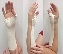
Figure 7 - Three Views of the Device after Application. Volar, Medial and Lateral Views. - Remove thermoplastic with two hands to avoid sticking, place it on the towel. Ensure it is orientated the way you would place it on the client.
- Roll distal edges at the DPC. Keep pattern nearby as a reminder of the correct side to roll. Do not roll at IPJ (Refer to Figure 6).

Application of thermoplastic to the client
- Return template into the water. Ensure patient is ready. Position supported forearm vertically. Using a goniometer, position wrist between 20-30 degrees extension, and thumb opposed to allow contact with index finger. Other fingers should be gently extended to ease application. Sorbolene cream can be used at thumb component to aid removal.
- Remove thermoplastic from water. Place on towel and allow to cool slightly before application. Test temperature of thermoplastic on client.
- Align thermoplastic on client. Ensure placement of distal edge is inferior to the DPC, equal width on lateral and medial sides along full length of forearm.

Figure 8 and 9 - Final Device. Lateral, Medial, Dorsal and Volar Views - Secure the thermoplastic to contours of the hand and forearm. Secure radial side first, followed by the thumb piece (loosely to aid removal).
- Check positioning again, ensuring optimal wrist extension. Form an ‘o’ with the thumb and index finger to obtain opposition.
- Use hands to mold and maintain ideal position whilst smoothing the plastic to avoid air pockets.
- Mark medial, lateral and proximal trimlines. Mark IPJ. Remove from client.

Finishing your device
- Cut along medial, lateral and proximal trimlines.
- Cut around IPJ. Dip this into the water until relatively soft. Roll at IPJ to ensure minimal restriction. Ensure this trimline is inferior to the IPJ on your client.
- Place proximal end of device into water at a 40 degree angle, until relatively soft. Flare bottom of the device with your palm. This minimises abrupt pressure changes.
- Return device onto the client to ensure ideal functioning. If particular aspect is not correct, return to water and repeat steps above.
- See Figure 7 for visual representation.
Strapping
- Measure length of strapping with device on the client, at three points – dorsal side of the MCPJ, the RCJ and proximal edge.
- Mark optimal positioning of strapping on device at each level.
- MCPJ Strap – Using a heat gun, permanently secure Velcro loop strap to radial side of the device by heating both the strap and the device. Press firmly. For the ulnar side, heat and attach a piece of hook velcro. Ensure hook and loop are aligned.
- RCJ and Proximal Strap – Heat both the device and hook/s, one at a time. Secure hook/s to device at marked positions. At the RCJ, the loop strapping attaches to same hook piece. Two hook pieces positioned on an angle will be required for the proximal strap to allow contour to the forearm. At both levels there is no fixed strapping. (See Figure 9 for positioning).
- Review strap placement and thickness. If pinching, bunching or pressure points develop, adjustment may be required. Alternatively, soft padding may be incorporated into strapping to relieve pressure areas.
- Refer to Figures 8 and 9 for attachment methods and final presentation of device.
Plaster of Paris Cast
[edit | edit source]Prepare the environment around you

- Ensure adequate space for you and your client. Client should be seated with elbow supported, ensure there is enough space to access the limb from all sides.
- Set up work area and materials, including rolls of Plaster and Paris, scissors, indelible pencil, bucket of water, stockinet, protective plastic sheets.
Prepare your pattern.
- Follow the instructions listed for LTT.
Prepare your client
- Place stockinet over client’s arm. Cut a small hole to allow thumb to exit. Secure a knot at the end of the stockinet to prevent slipping.
- Mark boney prominences and trimlines from the template with the indelible pencil.
- Apply Sorbolene and padding around thumb as required to aid removal of cast.
Casting

- Prepare your plaster. Measure plaster against the length of client’s arm. Allow 6-8 layers of plaster. Check to see if width of plaster exceeds trimlines, two slabs may be required. Measure and cut a hole for the thumb, allowing enough plaster to meet trimline at the DPC. Measure a small 6-8 layer piece of plaster for contained thumb component.
- Place client in appropriate position, as mentioned previously - supported forearm vertical, wrist between 20-30 degrees extension, thumb opposed and in contact with index finger, other fingers gently extended.
- Place the plaster into water, remove excess.
- Align plaster to client. Ensure placement of distal edge inferior to the DPC, equal width on lateral and medial sides along full length of forearm.
- Secure plaster to contours of the hand and forearm with long strokes. Small amounts of water can be applied to aid application.
- Align and secure thumb component ensuring a smooth transition between pieces. Using the indelible pencil, mark lines across the transition in case separation occurs.
- Position for optimal wrist extension. Form an ‘o’ with the thumb and index finger to obtain opposition.
- Wait until plaster goes off. Mark medial, lateral and proximal trimlines. Mark IPJ. Remove from client.
Finishing your cast
- Cut along medial, lateral and proximal trimlines, as well as the IPJ trimline.
- Review cast. Repeat above steps if needed.
- Refer to Figures 10 and 11 for visual representation of completed cast.
Critique of Fit
[edit | edit source]The Client
[edit | edit source]The client is a 7 year old girl who suffers from Spastic Cerebral Palsy. She presented with moderate wrist contracture in her right upper extremity, her dominant hand.
Subjective Assessment
[edit | edit source]On presentation, the client (with the assistance of her parents) reported that she has been receiving Botulinum Toxin A injections for approximately 3 months, to improve tone and range of motion. Although this treatment has shown significant improvements, the client expressed her frustration with her condition, as she is still unable to do the things she loves with ease. The client aims to be able to increase use of her right hand, for example, during play, writing and colouring, and daily tasks.
Objective Assessment
[edit | edit source]A physical assessment was completed and revealed
- At rest, the wrist is in a flexed position at 50°, with full thumb adduction. On manipulation, both the wrist and thumb can reach a neutral position. During the assessment, both active and passive range of motion of the wrist and thumb were tested. Both were between 10-20 degrees lower than normal range, with passive range of motion slightly larger than active range of motion. The client was able to obtain the range of movement at these joints with gravity eliminated, as well as against gravity, however movement was slightly restricted with resistance.
- Overall, client’s range of motion is quite good, due to the client’s Botulinum Toxin A treatment plan. Fair functioning was evident, with relatively adequate voluntary motor control.
Orthotic Goals
[edit | edit source]The goal of orthotic management for this client is to prevent further contracture of the wrist with the use of an orthotic device between Botulinum Toxin A treatments. The device will be in a functional position to allow the client to participate in various daily activities.
Prescription
[edit | edit source]- In order to achieve these goals, a low temperature thermoplastic Wrist Thumb Orthosis was prescribed. This device aims to immobilise the wrist in a neutral position with slight extension to support the phalanges in a functional position, with the CMC and MCPJ in an opposed position providing a stable pole for opposition. This will assist the client complete the various daily bimanual activities she hopes to achieve, for example, picking up and utilising objects efficiently.
- Literature suggests that a greater degree of wrist control is needed in patients with high tone and presence of contracture. As a result, Wilton (2003) suggests a more rigid functional splint is more appropriate. As mentioned by Feldman (1990), a rigid low temperature plastic is commonly used in the splinting of spastic upper extremities. By stabilising the wrist in a neutral position with a rigid functional low thermoplastic splint, muscular forces will transfer to the distal regions more efficiently (Wilton, 2003). A study by Louwers et al. (2011) investigated the immediate effects of the use of a static wrist and thumb brace on bimanual activities. Results indicated that static bracing of the wrist and thumb in a neutral position can improve performance in bimanual activities.
- Based on the above, the following device design was utilised.
Device Design and Final Presentation
[edit | edit source]- Positioning of the device includes the RCJ immobilised at 25 degrees extension, obtaining a relatively neutral position. The MCPJ of the thumb is immobilised in an opposed position, allowing contact with index finger.
- The device covers the volar surface of the limb, with the proximal trimline extending from 2/3 of the forearm to the distal trimline inferior to the Distal Palmar Crease. The distal trimline at the MCPJ extends inferior to the IPJ of the thumb. Medial and lateral trimlines extend to half the circumference of the forearm, with the medial trimline inferior to the ulnar styloid and lateral trimline inferior to radial styloid.
- Attachment methods included strapping over dorsal side of the MCPJ, the RCJ and proximal edge of the device. MCPJ Strap utilises a 2 cm Velcro, permanently secured to radial side of the device, with the RCJ and proximal strap utilising a 4 cm Velcro, with no permanent strapping.
- The immobilisation of the RCJ requires a downward manipulatory force at the RCJ, with two stabilising forces at the MCPJ and the proximal edge. The immobilisation of the MCPJ requires an inward manipulatory force at the MCPJ of the Thumb with two stabilising forces at the CMC and the IP joint of the thumb. These forces are achieved through the three attachment methods utilised.
Fit of the Device
[edit | edit source]- The overall technical standard of the device is fair, with neat and smooth trimlines, and an overall even surface contouring to the limb. However, there are a few areas which need improvement.
- When the client is in full elbow flexion, there is pinching evident at the proximal edge on the lateral corner (See Figure 12). Over time this may cause a pressure point and discomfort for the client. A greater degree of flaring could have been incorporated into the proximal edge of the device to prevent this.
- When the device is fitted to the client, there is a large amount of gaping at the thumb webspace (See Figure 13). During the application process, a lack of conformity to the limb within the webspace was evident. In future, more care would be taken to ensure adequate securing of the device to the contours of the client’s thumb and webspace.
- Flexion at the MCPJs causes the distal trimline of the device to obtrude slightly inferior to the 2nd digit, creating pressure at the point of contact. (See Figure 14). This is caused by an uneven trimline at the distal palmar crease, with a slight increase in height at this point obstructing MCPJ flexion. Further rolling of the distal trimline may prevent this from occurring.
- Flexion at the IPJ of the thumb causes pressure superior and inferior to the joint, further resulting in increased pressure at the CMC joint (See Figure 15). The trimline at the IP joint appears to be too high, restricting IP flexion. With further rolling at the IP joint, there may have been less pressure and restriction. If pressure at the CMC is still evident with a lower trimline present, a softer material or padding could be added across the CMC prior to the application of the device to relieve some pressure from the area.
- With prolonged use, the client felt increasing pressure and slight pain at Radial styloid (See Figure 16). This pain was felt more in some functional activities, including colouring. To reduce these symptoms, a lower trimline may be needed to reduce the pressure at this area. Alternatively, the edge could be flared slightly to reduce the sudden pressure change.
 |
 |
 |
 |
 |
Alternative Design
[edit | edit source]- Immobilisation of the MCPJ of the thumb, although in an opposed position, found to be limiting or restrictive when performing activities that utilised greater thumb abduction than the device allowed. A Wrist Hand Orthosis, whilst still immobilising the RC joint, may provide increased range of motion at the thumb to allow optimal functioning. However, as the client’s thumb presents in an abducted position, care and considerations would have to be made to avoid excessive pressure on the palmar surface at the CMC.
- Alternatively, a Wrist Hand Finger Orthosis may be beneficial as a resting orthosis for the client, to further aid the prevention of contracture whilst the client sleeps.
Demonstration of Device with Outcome Measures
[edit | edit source]- The Upper Extremity Functional Index (UEFI) was utilised pre and post treatment as an outcome measure to obtain an indication of functional benefits of the orthosis prescribed. Five items were excluded from the assessment, as they were not applicable to the client, for example, driving and ironing clothes. Because of this, a final score cannot be derived with integrity. However, we can compare the scores of individual items to assess improvements in a more focused sense. Score improvements can be seen in 13 of the 15 items, however, it is hard to determine if these improvements are significant.
- To compliment these results, the Patient-Specific Functional Scale (PSFS) was also used to explore functional changes in five important activities, as depicted by the client. An initial total score of 4.2 was achieved, with post treatment revealing a score of 7.2, indicating a significant positive change in function post treatment. Furthermore, items 1-4 were 3 points higher at the post treatment assessment compared to the pre-treatment scores. From this, we can conclude that a detectable positive functional chnage was achieved with the use of the orthotic treatment prescribed in both individual and overall scores.
- A more detailed outline and visual representation of the outcome measures performed can be viewed in the following section.
Outcome Measures
[edit | edit source]In order to assess and quantify positive functional changes as a result of orthotic treatment, two outcome measures were utilised, including the Upper Extremity Functional Index and the Patient-Specific Functional Scale. These two measures are specified by the Transport Accident Commission (2014) as standard outcome measures for health professionals to assist clinical practice.
The Upper Extremity Functional Index (UEFI) was utilised pre-treatment and 4 weeks post-treatment to obtain an indication of functional benefits of the orthosis prescribed. As one of the client goals was to increase functional use of her right hand, this measure was chosen as it provided a wide range of daily tasks that could evaluate and assess functional improvement.
The UEFI includes 20 items, producing a score out of 80, with 9 points being a minimum level of detectable change, as suggested by Stratford, Binkley and Stratford (2001). Five items were excluded from the assessment as they were not applicable to the client. Examples of these items include driving, as well as ironing clothes. Because five items were not applicable to the client, a score out of 80 cannot be derived. A total score out of 60 was utilised.
For this client, the total score for the pre-treatment assessment was 12 out of 60, with post-treatment generating a score of 32 out of 60. With the difference exceeding 9 points, a significant change may be evident. However, as some questions were excluded from the assessment, the maximum possible score will differ, and therefore the reliability and validity of the measure may be compromised.
Although a total score may not be an effective measure, we can also compare the scores of individual items to determine if there is any improvement in a more focused sense. Table 1 depicts the individual scores of the items utilised, pre- and post-treatment. Score improvements can be seen in 13 of the 15 items. However, as Stratford et al. (2001) do not provide a minimum detectable change for single activities, we are unable to determine if the change evident is a significant improvement.
 |
 |
 |
 |
 |
To complement the results of the UEFI, the Patient-Specific Functional Scale (PSFS) was also used to explore functional changes in five activities, as depicted by the client. This measure was selected as it allowed the client to choose five activities that were particularly important and performed on a daily basis. The five items included were colouring, tying shoe laces, opening a packet of chips, picking up small objects (eg. blocks or toys), and brushing hair. A visual representation of items 1-4 can be seen in Figures 17 to 20.
Developed by Stratford, Gill, Westaway and Binkley (1995), the PSFS allows a valid and reliable assessment of changes across the five individual items, as well as an overall score, for initial and follow-up assessments. The scoring scheme ranges from 0 to 10, with zero indicating the client is unable to perform the activity, and 10 indicating the client is able to perform activity at the same level as before injury or problem. A score of 10 in this case was utilised to describe the client being able to perform the activity overall. The minimum detectable change for the total average score is 2 points, with single item scores having a minimal detectable change of 3 points.
For the PSFS, a total score of 4.2 was achieved for pre-treatment, with a score of 7.2 for the post-treatment. Furthermore, items 1-4 were all three points higher at the post-treatment assessment compared to the pre-treatment scores. From these results, we can conclude that a detectable positive functional change was achieved with the use of the orthotic treatment prescribed.
The UEFI and the PSFS were utilised to evaluate positive functional changes resulting from the prescribed orthotic treatment. The UEFI provided a broad assessment of various daily activities, with the PSFS allowing a focus on five of the client’s common daily activities. Both measures were easy to administer, however the UEFI was quite a long measure and required a number of breaks to maintain the client’s attention. Although the UEFI provided a thorough evaluation, the PSFS alone may be a more suitable assessment for this particular client. In summary, the outcome measures utilised with this client revealed a positive change in function with orthosis use.
Referral Letter
[edit | edit source]20th of May, 2014
Susan Miller
ABC Physiotherapy
123 Middle St
North Melbourne
Dear Susan,
I am referring the current client as she would benefit greatly from regular physiotherapy. I hope that with the use of exercise and massage we can prevent further development of the contracture, improve and maintain existing benefits from the client’s Botulinum Toxin A treatment plan, and increase strength and flexibility of the muscles of the hand and forearm to optimise physical functioning.
The client is a 7 year old female who suffers from Spastic Cerebral Palsy. The patient presented with moderate wrist contracture in her right upper extremity, her dominant hand. As previously mentioned, the patient has been receiving Botulinum Toxin A injections for approximately three months to improve tone and range of motion.
At rest, the client’s wrist is in a flexed position at 50°, with full thumb adduction. On manipulation, the wrist and thumb can reach a neutral position. Due to the clients Botulinum Toxin A treatment plan, the client’s range of motion is quite good. Wrist flexion and extension is slightly lower than normal range. The client has relatively fair functioning, with adequate voluntary motor control, intellectual capacity and minimal sensory impairment.
In order to compliment the Botulinum Toxin A treatment, I have designed and manufactured a device which aims to aid the prevention of further contracture of the wrist. It is a functional device, with the wrist and thumb immobilised in a position which allows the client to participate in various daily activities.
The client has been wearing the device for approximately 4 weeks, with significant functional benefits developing in this time. For example, the use of the Patient-Specific Functional Scale demonstrated a detectable positive functional change in five specific activities with the use of the orthotic treatment prescribed in both individual and overall scores. These activities were deemed important to the client and performed daily, including brushing hair, colouring, and tying shoe laces. This improvement has been greatly appreciated by the client, with the opportunity for further improvement with your assistance.
I believe the inclusion of physiotherapy into the client’s current treatment program will be highly beneficial, focusing particularly on exercises that will improve and facilitate the client’s functional use of her right hand.
If you have any questions or concerns regarding the above information, please don’t hesitate to contact me.
Kind regards,
Cassandra Michal
Latrobe Prosthetics and Orthotics
123 Plenty Road
Bundoora
Search Strategy
[edit | edit source]For the research within this paper, the databases utilised included Cinahl, Medline and Pubmed. The key terms employed within this search included: Cerebral Palsy, Spasticity, Orthotic Management, Splint*, Upper Limb/Extremity, Effectiveness, Botulinum toxin A. Further articles were sourced from research mentioned within articles reviewed. The Cerebral Palsy Australia website was also utilised.
Reference List
[edit | edit source]Burtner, P. A., Poole, J. L., Torres, T., Medora, A. M., Abeyta, R., Keene, J & Qualls, C. (2008). Effect of wrist hand splints on grip, pinch, manual dexterity, and muscle activation in children with spastic hemiplegia: A preliminary study. Journal of Hand Therapy, 21, 36-43. doi: 10.1197/j.jht.2007.08.018
Cerebral Palsy Australia. (2010). What is Cerebral Palsy? Retrieved from http://www.cpaustralia.com.au/index.php/site/learningcentre/thefacts/whatiscp
Cerebral Palsy Australia. (2010) Types of Cerebral Palsy. Retrieved from http://www.cpaustralia.com.au/index.php/site/learningcentre/thefacts/typesofcp
Chin, T. Y. P., Duncan, J. A., Johnstone, B. R., & Graham, H. K. (2005). Management of the upper limb in cerebral palsy. Journal of Pediatric Orthopaedics, 14(6), 389-404. Retrieved from http://0-ovidsp.uk.ovid.com.alpha2.latrobe.edu.au/sp-3.11.0a/ovidweb.cgi
Dahlin, L. B., Komoto-Tufvesson, Y. & Saleback, S. (1998). Surgery of the spastic hand in cerebral palsy. Journal of Hand Surgery, 23(3), 334-339. doi: 10.1016/S0266-7681(98)80053-3
Feldman, P. (1990). Upper extremity splinting and casting. In M. B. Glenn & J. Whyte (Eds.), The practical management of spasticity in children and adults (pp.149-166). Malvern, PA: Lea & Febiger.
Hoare, B. J. & Russo, R. N. Botulinum toxin-A in children with cerebral palsy: Treatment of the upper limb following injection. In: Soderback, I editor(s). International Handbook of Occupational Therapy Interventions. New York: Springer Publishers, 2009. doi: 10.1007/978-0-387-75424-6-35
Hoare, B. J., Wallen, M. A., Imms, C., Villanuev, E., Rawicki, H. B. & Carey, L. (2010). Botulinum toxin A as an adjunct to treatment in the management of the upper limb in children with cerebral palsy. Cochrane Database of Systematic Reviews, 1, 1-97. doi: 10.1002/14651858.CD003469.pub4
Imms, C. (2011). Bracing and splinting interventions in the upper limbs of people with cerebral palsy. Developmental Medicine & Child Neurology, 53(4), 293-294. doi: 10.1111/j.1469-8749.2010.03881.x
Kanellopoulos, A. D., Mavrogenis, A. F., Mitsiokapa, E. A., Panagopoulos, D., Skouteli, H., Vrettos, G., Tzanos, G., & Papagelopoulos, P. J. (2009). Long lasting benefits following the combination of static night upper extremity splinting with botulinum toxin A injections in cerebral palsy children. European Journal of Physical and Rehabilitation Medicine, 45(4), 501-506. Retrieved from http://0-search.proquest.com.alpha2.latrobe.edu.au/docview/777808334
Lannin, N A., Cusick, A., McCluskey, A. & Herbert, R. D. (2007). Effects of splinting on wrist contracture after stroke: A randomize controlled trial. Stroke, 38(1), 111-116. doi: 10.1161/01.STR.0000251722.77088.12
Lannin, N. A., Novak, I., & Cusick, A. (2007). A systematic review of upper extremity casting for children and adults with central nervous system motor disorders. Clinical Rehabilitation, 21(11), 963-976. doi: 10.1177/0269215507079141
Louwers, A., Meester-Delver, A., Folmer, K., Nollet, F. & Beelen, A. (2011). Immediate effect of a wrist and thumb brace on bimanual activities in children with hemiplegic cerebral palsy. Developmental Medicine & Child Neurology, 53(4), 321-326. doi: 10.1111/j.1469-8749.2010.03849.x
Stratford, P. W., Binkley, J. M., & Stratford, D. M. (2001). Development and initial validation of the upper extremity functional index. Physiotherapy Canada, 53(4), 259-267.
Stratford, P., Gill, C., Westaway, M., & Binkley, J. (1995). Assessing disability and change on individual patients: a report of a patient specific measure. Physiotherapy Canada, 47, 258-263.
Teplicky, R., Law, M. & Russel, D. (2002). The effectiveness of casts, orthoses, and splints for children with neurological disorders. Infants and Young Children, 15(1), 42-50. doi: 00001163-200207000-00007
Transport Accident Commission (2014). Standard Outcome Measures. Retrieved from https://www.tac.vic.gov.au/providers/clinical-resources/outcome-measures#ul
Wilton, J. (2003). Casting, splinting, and physical and occupational therapy of hand deformity and dysfunction in cerebral palsy. Hand Clinics, 19(4), 573-584. doi: 10.1016/S0749-0712(03)00044-1
Yang, T. F., Fu, C. P., Kao, N. T., Chan, R. C., Chen, S. J. (2003). Effect of botulinum toxin type A on cerebral palsy with upper limb spasticity. American Journal of Physical Medicine and Rehabilitation, 82(4), 284-289. doi: 10.1097/01.PHM.0000056989.67763.07