The patient is a 42 year old female who was diagnosed with rheumatoid arthritis (RA) 4 years earlier. She runs her own business as a jewellry designer and manufacturer and as such does a lot of work with her hands. Although she first presented with RA in both of her knees she has recently begun to have pain and inflammation in her wrists particularly at the end of the working week. There are no signs of ulnar drift in the wrists as a result of the RA at present.
Due to a stomach ulcer when she was younger she is unable to take NSAIDs due to the contraindications of the drugs. Rest, activity avoidance and occasionally heat therapy are providing her with some limited relief.
The patient would like to be able to continue working however the pain and stiffness she is experiencing each day in her wrists is limiting her.
Orthotic Treatment of Rheumatoid Arthritis of the Wrist
An autoimmune disease, rheumatoid arthritis (RA) is a devastating disease that typically affects adults between 20 and 40 years of age [1][2]. Although its aetiology is in part still unknown, what is known however, is that women are two to three times more likely to be afflicted than men, with genetic involvement contributing to the risk of contracting the disease [1][3][2]. A systemic polyarticular disease, RA occurs symmetrically most often in the joints of the feet, ankles, hands, wrists and knees [4][1][5]. The destruction of the joints begins with persistent synovitis progressing through to irreparable damage to articular cartilage, ligaments, tendons and the underlying bone [1][2]. The formation of excess granulation tissue in the form of a Pannus often results in the disabling deformities often associated with the disease [1][2]. Synovial hypertrophy alters the normal biomechanics of the wrist and hands often resulting in the wrist collapsing into radial deviation with ulnar drift of the fingers [5] and/or boutonniere and swan neck deformities [4]. Symptoms of RA can include fatigue, joint swelling, decreased muscle strength and most importantly pain [6]. No two patients will follow the same disease progression, with periodic flares and remissions of the disease commonplace [4]. Fortunately treatment for RA can be successful in slowing the disease progression, delaying the disabling effects and may consist of conservative and pharmacological management techniques [1][5]. An orthosis is one such example of conservative treatment routinely provided to persons with RA.
Rheumatoid joint capsule
An orthosis is a rigid or semi-rigid device used to provide alignment and/or support, improve function, correct or prevent deformity or restrict movement of a body part [7][5]. For persons with RA, devices that can provide strength, comfort and protection are often custom made but are also available prefabricated, sometimes in a range of different materials to suit individual needs [8][7]. Ideally the orthosis should be easy to donn and doff with modifications to straps accommodating for the limited dexterity that may be affecting the patient [4]. Long-term use of corticosteroids often prescribed for patients with RA can also leave their skin thin, delicate and able to bruise more easily consequently, providing padding over bony prominences and eliminating areas that may cause friction are essential [9][4][7]. Although RA commonly affects the hands and elbows, the wrist complex is estimated to affect up to 75% of persons with the disease [3][10] resulting in loss of hand function, grip strength and dexterity [7][10].
The wrist complex is comprised of the radiocarpal and midcarpal joints, both of which are surrounded by a joint capsule lined with a synovial membrane and strengthened medially by the ulnar collateral ligament and laterally by the radial collateral ligament [11]. The radiocarpal joint consists of the distal radius and the lunate and scaphoid. The remaining carpal bones, articulating with the proximal surfaces of the lunate scaphoid and triquetrum make up the midcarpal joint [11]. Blood supply to the complex is via branches of the dorsal and palmar arches with innervation via branches of the ulnar, medial and radial nerves [11]. There are nine muscles located in the forearm the tendons of which cross the wrist and are inserted in the hand that work together to produce the various movements possible at the wrist. Extension of the wrist is produced by the extensor carpi radialis longus, radialis brevis and extensor carpi ulnaris [12][11]. Flexion is possible due to the flexor carpi ulnaris and flexor carpi radialis muscles with some assistance from the finger and thumb flexors and palmaris longus [12][11]. Adduction of the wrist occurs when extensor carpi ulnaris and flexor carpi ulnaris work together whilst abduction requires the combination of flexor carpi radialis, extensor carpi radialis brevis and longus along with abductor pollicis longus [11]. With its combination of complex articular surfaces, muscular, vascular and nerve supplies, any injury or inflammation at this joint complex can affect movements in other joints both proximally and distally. The importance of preserving the functionality of these joints should not be underestimated. In order to function optimally, joints of the upper limb need to be able to move smoothly and friction free, their small cartilaginous articular surfaces unhindered by scars or adhesions that may restrict their movement[4]. In particular, the functioning of the hand may be reduced significantly if there is anatomic re-arrangement of tissues or an alteration to its physical characteristics[4]. Unfortunately, due to the very nature of the disease, RA frequently alters the alignment and positioning of affected joints, particularly in its advanced stages[1][2].
There are numerous treatment options available to persons with RA of the wrist including various drug treatments, steroid injections, surgical intervention and assistive devices. For the purposes of this discussion, only the orthotic options available to persons with RA of the wrist will be explored and compared. Orthoses utilized by persons with RA of the wrist can be placed into three main categories; immobilising, mobilising and postoperative [4]. Immobilisation splints also referred to as ‘resting‘ orthoses are recommended for use during acute periods of inflammation, alleviating the pain caused by the inflamed tissues and can be prefabricated or custom made [4][13]. Mobilisation orthoses occasionally use elastic traction to aid in joint positioning and minimize joint contractures [4][7]. They can also be known as ‘working’ orthoses as the designs allow the user to continue with their activities of daily living with very little restriction [4][13][7]. Some of the prefabricated ‘working splints’ available consist of a fabric gauntlet with a removable volar metal stay, their designs similar, with placement of the straps often differing [10]. Postoperative orthoses as their name suggests are used after a patient has undergone surgery, providing protection and immobilisation to facilitate healing [4]. The design must take the condition of the soft tissues and surgical incisions into consideration and close communication between the surgeon and the team of allied professionals is essential for a positive outcome [4][7].
Research into the value and efficacy of these devices has been sporadic and of mixed opinions. Two Cochrane reviews have been conducted into the use of these orthoses, one performed by Egan and colleagues (2001)[14] and the other by Steultjens and her colleagues (2004)[6] which specifically discussed rheumatoid arthritis treatments from the Occupational Therapists’ perspective. The first review came to the conclusion that no evidence supporting the use of either resting or working splints to reduce pain or improve dexterity existed, at that time [14]. Interestingly, the second review highlighted studies that advocated the use of orthoses for pain reduction and improvement of grip strength, although these gains came at the cost of dexterity [6]. In the 10 years that have passed since these reviews were conducted, further studies have been completed of varying quality and mixed conclusions. A randomised control trial performed in 2008 collected data that suggested that static resting orthoses are of no benefit[15]. Upon closer inspection however, it is clear that this study was flawed due to low compliance rates and recruitment of subjects who did not even have a confirmed diagnosis of RA [15]. Another study in 2008 compared three different ‘working’ orthoses, 2 prefabricated and 1 custom made and indicated that use of the devices reduced pain significantly and improved grip strength [8]. Similarly, Veerhof and colleagues in 2008 also investigated working orthoses and likewise concluded that pain relief was evident after 4 weeks of use[10]. Although these studies were more robust than that of Adams et al. (2008)[15], what is evident is that more research is warranted into the effectiveness of orthoses for the treatment of RA of the wrist.
It is hoped that by providing a resting orthosis for a patient with RA of the wrists, the various muscular, bony and ligamentous components of the wrists could ‘rest’ during periods of disease activity. That is, they would be able to maintain correct alignment, pain producing movement would be restricted and an opportunity for the inflammation to heal without further aggravation would be provided. In particular, the distal radius, carpals, metacarpals, and phalanges would need to be immobilised. It would also be important to ensure that pressure applied by the orthosis is well distributed over the area in contact with the skin. The straps should be easy to apply and remove, to allow for the limited dexterity patients with RA must often contend with.
Due to the characteristic flares and remissions of symptoms common to patients with RA [2], particularly in the early stages, the resting orthosis could be worn on an ‘as need’ basis during periods of symptomatic flare ups.
By providing this relief, it is hoped that the patient would be able to return to work and be far more productive as a result of the reduction in pain.
Although a cast made from POP bandages was taken and might be used to make a definitive device from a high temperature thermoplastic (HTT), the additional time required to make the device along with the potential discomfort to the client would not be warranted, particularly when the device would still only be worn on an ‘as need’ basis as their symptoms increase.
Since RA is a disease that affects joints bilaterally, a resting orthosis would ideally be manufactured for both the left and right sides. For the purposes of this project only one for use on the left forearm and hand will be described and manufactured.
The nature of the patient’s vocation requires her to perform her work using fine motor skills. Utilisation of a ‘working orthosis’ would be counter-productive, particularly in light of the lack of research supporting their use. Instead, provision of a pair of resting orthoses that the patient could wear at night in order to relieve discomfort and prevent joint deformity would be the recommendation of this researcher. There is a tendency for the metacarpophalangeal (MCP) joints to sublux palmarly, therefore an orthosis fitted to the volar aspect of the forearm and hand would help to support these joints [4][13][7]. Ideally the MCP joints would be in 25-30 degrees of flexion to provide palmar support to these joints and their surrounding tissues [4]. Additionally, the interphalangeal joints would be in 30 degrees of flexion and the wrist in 0-10 degrees of flexion as too much flexion might put pressure on the carpal tunnel that is likely to already be compressed due to the inflammation of the wrist joint [4][7]. Additionally the wrist should be neutral for rotation, ulnar and radial deviation. The thumb would also ideally be placed in palmar abduction, again to reduce pressure on the surrounding and underlying tissues [4].
The trimlines of the orthosis should ideally be contoured in such a way that they do not pinch or ‘dig into’ the soft tissues of the patient’s limb. The length of the orthosis should be 2/3 the length of the forearm and the medial and lateral walls should come half way up the sides of the forearm. The proximal edge of the orthosis should be flared to protect the soft tissues.
Due to the often limited dexterity of a person with RA (caused by the inflammation, pain and sometimes joint deformities), the straps would have small holes of approximately 1cm diameter cut into them in order to facilitate easier removal.
The straps would be located across the dorsal surface of forearm at the proximal end, across the dorsal surface of the wrist joint and across the dorsal aspect of the interphalangeal joints. These straps would all be completely removable if necessary. A small strap for the thumb would be permanently adhered on the medial side and would wrap around the thumb attaching on the lateral side.
Padding made from a lightweight, soft and durable foam such as Pelite is added to the straps over the bony prominences of the interphalangeal joints and the ulnar styloid process to reduce the pressure over them. A second set of straps are provided without padding during times of extreme inflammation and swelling when the padded strap might feel too tight. The ends of the straps should be rounded using scissors to create a more attractive cosmetic appearance.
Sticky-back Velcro loop should be used to adhere the straps to the LTT and Velcro hook for the straps themselves, cutting both parts the same width so that maximum adherence can be achieved.
• Materials required – Chair, table, pen, measuring tape or ruler, scissors, kitchen cloth to make pattern, piece of LTT, electric frypan, water, goniometer, sticky back Velcro loop and Velcro hook, sandpaper (80-180 grade)
• In order to create the pattern needed for the resting orthosis use a kitchen cloth such as a 'Chux' large enough to rest the patients hand and forearm. Lay it flat on a table top or other suitable flat surface. Paper underneath might be needed as the texta may come through the cloth.
• With the patient seated ask them to rest their forearm on the cloth with their palmar surface on the cloth (Fig 1). Keeping their digits reasonably close together in a relaxed position trace around the forearm and hand (Fig 2). Make a mark at the base of the CMC joint just distal to the radial styloid process (Fig 3).
• At a point 40mm or two finger widths wide distal to the cubital fossa, make a mark on the cloth on the lateral and medial sides of the forearm as this mark gives the length required.
• Using a texta or marker draw a new pattern onto the chux that is the same shape as the original but with a seam allowance on the lateral and medial sides of the forearm. Roughly 2cms allowance should be enough as the final trim lines will be determined once the orthosis has been formed. The seam allowance is only needed to extend up to the base of the palm on the ulnar side and just distal to the tip of the thumb on the radial side (Fig 3)
• Measure the length of the thumb placing the ruler into the webspace. (For clarity this will be known as Measurement A) (Fig 4). Add two centimetres to this measurement.(Measurement B) (eg, Thumb measurement A = 7cm, new measurement B = 9cm) Halve measurement B (eg. 4.5cm) and locate this point on the ruler. Line this point on the ruler up with the CMC mark made on the original tracing. With the ruler in place, mark both ends of measurement B onto the original tracing line (Fig 5) These marks indicate the length of the inverted thumb piece of the orthosis. Round the edge of the most inferior mark out to the trimlines so that it resembles an inverted thumb.
• From the original traced line, draw a line on a 45 degree angle downwards to the new trimline edge. On the edge closest to the thumb cut a ‘u shaped’ curve into the chux to allow for the thenar eminence when the thumb piece is formed. Be sure to cut the v-shaped gap between the thumb and the palm of the hand deep as the thumb piece will be twisted around slightly when formed.
• Cut the rest of the pattern out and apply to the patient’s arm to check for fit. Adjust trimlines if too excessive.
• When happy with the size and fit, trace the cloth pattern onto the low temperature thermoplastic (LTT) marking it with pen or another medium that can easily be cleaned off later. Cut the pattern out and when ready to mould, place in the water bath to soften (Fig 6)
• Whilst the LTT is softening, using a goniometer to check the angles on the patient's wrist, MCP and IP joints are correct (see design specifications) and ask them to hold them in the correct position (Fig 7)
• With the arm supinated place the warmed LTT over the forearm and hand being careful to ensure the thumb piece does not adhere to the other parts as it will be very prone to do this (Fig 8).
• There will be excess material along all of the edges which will be trimmed later. Ensure the positioning of the material is correct then work on moulding the LTT under the fingers, onto the thumb and around the wrist and forearm. The thumb piece needs to be lifted up and will rest on the top of the thumb in its abducted position.
• When the orthosis has cooled, mark the trimlines again (the material will stretch and so there will be excess to trim) then remove from the patient and trim excess. Fit again to the patient and check for comfort and alignment. Mark placement of straps whilst orthosis is on the patient. Prepare the velco loop pieces for the straps and hook attachment pieces. Padding in a light foam such as Pelite can be cut to size and the edges neatened as necessary. These pads should then be attached to the two most distal straps using an industrial sewing machine.
• Using a heat gun, warm the section of the orthosis where the Velcro hook needs to be attached then heat the sticky side of the hook piece, holding it with the ends of a pair of scissors. Carefully place the hook onto the LTT where required. Repeat for all pieces that require adhering. On the thumb of the orthosis gently warm a small section of the thermoplastic where the thumb strap needs to adhere. Warm the loop side of the Velcro strap cut for the thumb and whilst still hot press onto the warmed section of LTT on the thumb.
• Clean any remaining pen marks with acetone and ensure edges of the orthosis are smooth. If there are rough patches use a small piece of sandpaper (80-180 grade) to smooth them off.
• When all straps are finished, fit the patient with the orthosis and check for fit, making sure straps are the right length and adjust if necessary.
Straps and attachment placement on finished orthosis
The patient is a 42 year old female who lives in a single story house with her partner and two children. She works for herself as a jewellery designer and manufacturer.
The patient was diagnosed with Rheumatoid arthritis 4 years ago and at that time it only affected her knees. Since then it has progressed to her wrists and hands, worsening over the past 8 months. She was diagnosed with a stomach ulcer 15 years ago which has since healed but due to her susceptibility for this type of gastrointestinal problem and the chance of re-occurrence, she is unable to take any anti-inflammatory medications to help with the RA.
She presented to the clinic 2 weeks ago with a referral from her rheumatologist requesting we provide her with a pair of resting orthoses to wear at night as the swelling and inflammation from her RA at times becomes quite debilitating. The patient reported that recently the pain in her knees is generally quite minimal so no testing was performed on the patient’s knee joints on this occasion.
Patient assessment form
Objective Information
A physical examination of the patient was conducted with particular attention paid to the wrists and hands. Clearance tests on the lower limb were performed and cleared. ROM tests were performed on the wrists and although the ROM was within normal limits, the patient expressed discomfort whilst performing the tests, particularly wrist flexion and ulnar/radial deviation. Due to her discomfort, further testing of the phalanges was not carried out (Please refer to Patient Assessment form for further details)
Upon examination the 1st CMC and MCP joints of both hands were slightly swollen, red and warm to the touch. The patient reported that on this occasion her pain levels were quite low despite the visual signs indicating inflammation. No neurological or reflex tests were performed on this occasion.
As a baseline, outcome measures of the DASH (Disabilities of the Arm, Shoulder Hand) Questionnaire and the PSEQ (Pain Self Efficacy Questionnaire) were given to the patient to complete prior to provision of the orthotic devices. The same questionnaires will be given to the client again once a number of weeks of treatment have elapsed to use as a comparison.
Orthotic Goals and Prescription
Although there is currently no deformity present in any of her joints, preventing deformity is a very important functional aim, in addition to the reduction of inflammation, swelling and pain. As the symptoms of RA can come and go as flares and remissions, the resting orthoses provided would only be worn on an ‘as need’ basis, alternating hands each night. Comfort and practicality, in addition to ease of application and removal of the devices are also important considerations.
Device Critique
Although a resting orthosis for each wrist and hand would ordinarily be provided, only the left hand orthosis was made on this occasion.
The patient was very compliant throughout the manufacturing process. Fortunately her pain levels were low on the day of manufacture so she was able to hold the joint positions she was placed her in for the moulding of the LTT. Overall, the final result was a good representation of the original design with the trimlines and angles achieved meeting the design specifications. Application of the three straps meant that the forces required to help maintain the wrist and hand in the resting position were obtained and positioned correctly. The loops provide not only an aid for easy application and removal of the device, whilst considering dexterity limitations, but also a visual reminder of which direction the straps needed to be pulled on to be removed/applied correctly. Being fully removable the direction the straps need to be applied can be altered if the patient required it. The padding on the straps was well positioned for the patient and being sewn on will prevent the padding from slipping or falling off. :Additional straps without padding were also supplied in the event the patient preferred them. Although the overall result was very good, there were a problems associated with the manufacture that are worth highlighting.
LTT stretches as it is heated and unfortunately some of the LTT around the opening made for the thenar eminence stretched a little, although due to its location, this stretch does not affect the overall functionality or integrity of the device and is more of a cosmetic detraction. A small amount of green permanent marker used to mark trimlines remained on the LTT also despite best efforts to remove them, they remained but ultimately it is only a cosmetic problem (see picture below)
The trimlines were adjusted as per the design specifications except for the proximal trimline that was shortened slightly so that the LTT pattern could fit in the water bath. A larger water bath would be utilized in future to eliminate the need for pattern size reduction. The joint angles required as per the design specifications were all obtained after moulding also.
The position of the LTT over the patient’s limb was not optimal, draped more over the lateral side than the medial. The lateral edge next to the 5th digit is much higher than the medial side.
The index finger is overlapped by the middle finger due to that side being so cramped (see picture below). The patient complained that the pressure being applied to the lateral side of the DP joint of the little finger could potentially be quite painful over a long period of time wearing the device (see picture below).
The moulding under the fingers was likewise less than ideal and the patient reports that although it did not cause pain, over time it might prove to be annoying or irritating (See picture below).
A way to combat all of these moulding problems would be perhaps to take a little more time to position the LTT before moulding it into the patient’s anatomy in addition to making the entire digit support section flatter whilst still maintaining the functional angles required. This would probably make the orthosis more comfortable.
Moulding of the LTT onto the forearm was mostly well contoured to the shape of the patient’s arm but one section along the lateral side just proximal to the second strap has slight crease in it. This twist does not affect the functioning of the orthosis nor does it cause any discomfort to the patient.
After wearing the orthosis for a short time, the patient reported that a small section of the orthosis at the lateral side of the proximal pinched their skin a little if their elbow was fully flexed(see picture below).
This is likely due to the orthosis being moulded when the elbow was extended and supinated. As the elbow is flexed, the muscle bellies shorten making that section of the forearm bigger. Perhaps if the orthosis had been moulded with the arm flexed, this problem might have been avoided.
Stretched LTT and pen marks
Overlapping of digits
LTT inferior to digits overshaped
Crease produced when moulding
Pinching at proximal trimline
Before the patient is sent home with the device, modifications will be made to improve the comfort. An appointment will be scheduled with the patient to return in 4 weeks to check on the comfort and effectiveness of the devices.
A referral to see an occupational therapist has also been provided to the patient so that she can be given joint protection therapy as an adjunct to the orthotic management.
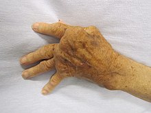
Without treatment, joint destruction that occurs as a consequence of Rheumatoid arthritis in the wrists and hands can lead to crippling deformities such as ulnar drift [4]. Ulnar drift as a result of Rheumatoid Arthritis It is therefore of paramount importance to prevent deformity as long as possible using means such as medication, orthotic intervention and joint protection.
The provision of a resting orthosis to reduce pain, inflammation and swelling and prevent deformity for this patient is appropriate as she uses her hands for her vocation. The outcome measures employed in order to justify the provision of the resting orthoses were the Disabilities of the Arm, Shoulder and Hand (DASH) [16] questionnaire and the Pain Self-Efficacy Questionnaire (PESQ)[17]. As the orthoses are utilised to reduce pain and inflammation, it seemed more pertinent to use outcome measures that could give an indication of improvement following rest whilst using the orthoses. Both measures are easily repeatable and valid measurement instruments.
The questionnaires were first taken prior to use of the resting orthoses. The second time the tests were taken was 4 weeks after the orthoses were provided and utilised. The PESQ test scores were obtained by adding the numerical responses to all of the questions together with a possible score of 60 points possible. The DASH scores were tabulated using the online calculator provided by the DASH Outcome Measure website with the responses manually entered onto the electronic version of the form [16].
Test Administered
PESQ (Pre)
PESQ (Post)
DASH (Pre)
DASH (Post)
DASH Work Module (Pre)
DASH Work Module (Post)
Score Obtained
23/60
35/60
58.5
40.0
81.3
56.3
As listed in the table above, there was an overall improvement in the PESQ score of 12 points. Since a 9 point increase in a PESQ score constitutes a minimal clinical important difference (MCID)[18], this result is a positive confirmation that the resting orthoses were successful in reducing the patient’s pain to a degree that improved their self-efficacy. The DASH scores (as listed above) likewise showed an improvement. The MCID for the DASH is 10.2 [19] although in this case the favourable outcome is a reduction in the score. The scores registered for both the regular section and the optional Work module reduced by 18.5 and 25 respectively. As is evident by the results listed above, use of the resting orthoses for a period of 4 weeks reduced pain and increased functional ability.
I would like to refer a patient of mine to you as I think your skills as a hand therapist could be of great help to her. Ms Boyden is a 42 year old who was diagnosed with rheumatoid arthritis 4 years ago. The disease first presented in her knees but is now also affecting her wrists and hands bilaterally. She experiences irregular flares of symptoms that can last from 1 week to 2 months or more. Thus far she has exhibited no signs of deformity and is currently not taking any medications for her condition. A previous gastrointestinal condition means she is unable to take anti-inflammatory medications. Ms Boyden works as a self-employed jeweller and as such has a great need for functional use of her hands. She has good active ROM in both hands within normal limits which is only restricted during inflammatory flares. I have manufactured a pair of resting orthoses for her to use at night when she is experiencing inflammation and its associated pain. It is hoped that the use of these orthoses will prevent joint deformity for as long as possible. I believe she could greatly benefit from joint protection therapy that will enable her to continue to carry out her activities of daily living and return to work in the short term and help prevent deformity in the long term.
If there is any further information you require pertaining to this patient please do not hesitate to contact me on 9688 4555 during business hours.
↑ 1.01.11.21.31.41.51.6Kumar, V., Abbas, A. K., Fausto, N., & Mitchell, R. N. (Eds.). (2007). Robbins basic pathology (8th ed.). Philadelphia, PA: Saunders/Elsevier.
↑ 2.02.12.22.32.42.5Scott, D. L., Wolfe, F., & Huizinga, T. W. J. (2010). Rheumatoid arthritis. The Lancet, 376(9746), 1094–108.
↑ 3.03.1Oldfield, V., & Felson, D. T. (2008). Exercise therapy and orthotic devices in rheumatoid arthritis: evidence-based review. Current Opinion in Rheumatology May 2008, 20(3), 353–359. doi:10.1097/BOR.0b013e3282fd17df
↑ 5.05.15.25.3Rizzo, M., & Cooney III, W. P. (2011). Current Concepts and Treatment for the Rheumatoid Wrist. Hand Clinics, 27(1), 57–72. doi:10.1016/j.hcl.2010.09.004
↑ 6.06.16.2Steultjens, E. E., Dekker, J. J., Bouter, L. M., Schaardenburg, D. D., Kuyk, M.-A. M., & Van den Ende, E. C. (2004). Occupational therapy for rheumatoid arthritis. In Cochrane Database of Systematic Reviews. John Wiley & Sons, Ltd. Retrieved from http://onlinelibrary.wiley.com/doi/10.1002/14651858.CD003114.pub2/abstract
↑ 7.07.17.27.37.47.57.67.77.8Jacobs, M. A., & Austin, N. M. (2013). Orthotic intervention for the hand and upper extremity: Splinting principles and process. Lippincott Williams & Wilkins.
↑ 8.08.1Haskett, S., Backman, C., Porter, B., Goyert, J., & Palejko, G. (2004). A crossover trial of custom-made and commercially available wrist splints in adults with inflammatory arthritis. Arthritis Care & Research, 51(5), 792–799. doi:10.1002/art.20699
↑Callinan, N. J., & Mathiowetz, V. (1996). Soft versus hard resting hand splints in rheumatoid arthritis: Pain relief, preference, and compliance. The American Journal of Occupational Therapy, 50(5), 347–353. doi:10.5014/ajot.50.5.347
↑ 10.010.110.210.3Veehof, M. M., Taal, E., Heijnsdijk-Rouwenhorst, L. M., & van de Laar, M. a. F. J. (2008). Efficacy of wrist working splints in patients with rheumatoid arthritis: A randomized controlled study. Arthritis Care & Research, 59(12), 1698–1704. doi:10.1002/art.24078
↑ 11.011.111.211.311.411.5Moore, K. L., Dalley, A. F., & Agur, A. M. R. (2010). Clinically oriented anatomy (6th ed.). Baltimore, M.D.: Lippincott, Williams and Wilkins.
↑ 12.012.1Bowker, P., Condie, D. N., Bader, D. L., & Pratt, D. J. (1993). Biomechanical basis of orthotic management. Oxford, UK: Butterworth-Heinemann.
↑ 13.013.113.2Hsu, J. D., Michael, J. W., & Fisk, J. R. (Eds.). (2008). AAOS atlas of orthoses and assistive devices (4th ed.). Philadelphia, PA: Mosby/Elsevier.
↑ 14.014.1Egan, M., Brosseau, L., Farmer, M., Ouimet, M.-A., Rees, S., Tugwell, P., & Wells, G. A. (2001). Splints and orthosis for treating rheumatoid arthritis. In Cochrane Database of Systematic Reviews. John Wiley & Sons, Ltd. Retrieved from http://onlinelibrary.wiley.com/doi/10.1002/14651858.CD004018/abstract
↑ 15.015.115.2Adams, J., Burridge, J., Mullee, M., Hammond, A., & Cooper, C. (2008). The clinical effectiveness of static resting splints in early rheumatoid arthritis: a randomized controlled trial. Rheumatology, 47(10), 1548–1553. doi:10.1093/rheumatology/ken292
↑ Di Pietro, F., Catley, M. J., McAuley, J. H., Parkitny, L., Maher, C. G., Costa, L. da C. M., … Moseley, G. L. (2014). Rasch Analysis Supports the Use of the Pain Self-Efficacy Questionnaire. Physical Therapy, 94(1), 91–100.
↑ Roy, J.-S., MacDermid, J. C., & Woodhouse, L. J. (2009). Measuring shoulder function: A systematic review of four questionnaires. Arthritis Care & Research, 61(5), 623–632. doi:10.1002/art.24396.
Searches were undertaken using the la Trobe University database in addition to the Medline, Cochrane and PubMed databases. Limits were placed on most of the searches so that articles were generally no more than 10 years old. All articles were additionally checked for relevance of their own references and used accordingly. Search terms used included "Rheumatoid arthritis", Orthos*, "orthotic*, "splint", "brace", "assistive device", "resting splint", "working splint".