
Upper Limb Orthotics/Partial Lesion of C7
Case study
[edit | edit source]The patient is a 70-year-old independent female who suffered a fall from a ladder resulting in a fracture in C7 4 months ago. Consequently, she experiences slight weakness when extending her wrist and lack of finger movement. Passive and resisted range of motion of her wrist and metacarpal-phalangeal joints were conducted to determine the strength of the movements based on the muscle strength grading scale. Her wrist extensor muscles were graded a 3 and her finger flexors were rated a 2. Additionally, strength was tested with lateral grasp (lateral pinch) with a pinch meter and palmar grasp (power grip) tests which the patient had difficulty performing. Neurological tests were conducted and passed Based on her motor function, C7 was affected. She was diagnosed with incomplete motor function with function below C7 intact. Due to poor management of her hand, she is unable to produce full extension of her wrist.
Evidence
[edit | edit source]Introduction
[edit | edit source]The spinal cord is part of an essential pathway in which the brain communicates with the body. The spinal cord contains white matter and neuron cell bodies known as gray matter that conduct a sensory or motor path (Kirshblum et al, 2011). A disruption to the pathway will interfere with function innervated by the affected spinal level, and a common mechanism of injury is blunt trauma such as motor vehicle accidents and impacts at high speeds. Injury to a spinal level can impact motor and sensory function can be exclusive to that level or include all the inferior levels. Tetraplegia or ‘Quadriplegia’ is often used to describe the loss of motor or sensory function in the cervical level, often a disruption in the spinal canal (Kirshblum et al, 2011) and results in loss of upper limb function. Similarly, brachial plexus lesions will impair upper limb function but differ from SCI due impairment limited to the upper limb and the differing mechanism of injury, which is due to forces acting on the shoulder and causing trauma to the network of nerves in the brachial plexus (Brunelli, 1991).
Classification
[edit | edit source]The American Spinal Injury Association (ASIA) devised a classification system for spinal cord injuries, categorising the level of impairment on a scale known as the ASIA Impairment Scale. Through conducting clinical assessments on suspected spinal cord injuries, the level of injury can be determined and whether sensory or motor function is impaired. To determine the motor level affected, manual muscle grading is utilised by determining what spinal level lacks key muscle activity. For C7, it is the triceps which enable elbow extension. If a level lacks the key muscle function, the rostral spinal nerves will increase in muscle grade strength (Kirshblum et al, 2011). Muscle grade is ranked 0-5, where 0= paralysis, 1= muscle contraction with no movement, 3= active full range of motion against gravity and 5= active full range of motion against gravity, with resistance.
Sensory examination is employed with “joint movement appreciation and position sense tests” (Kirshblum et al, 2011), which involve patients identifying the upper limb movement the examiner is producing whilst blinded and identifying the static position the upper limb is in. Alternatively, touch and pressure tests can be conducted.
A spinal cord injury can be described as complete, incomplete or normal based on the following characteristics:
Table 1: ASIA Impairment Scale
| Type | Characteristics |
|---|---|
| A | Complete: No sensory or motor function is preserved |
| B | Incomplete (Sensory): Sensory function preserved; motor function impaired |
| C | Motor function below spinal level is preserved OR more than half the major muscles below spinal level have a muscle strength grade below 3 |
| D | Incomplete (Motor): Motor function below spinal level is preserved; at least half major muscles below spinal cord have a muscle strength grade greater than 3. Voluntary anal sphincter contraction maintained. |
| E | Normal |
Anatomy and Physiology
[edit | edit source]Motor function derives from the ventral roots, which contain efferent nerve fibres that arise from neurons of the ventral horn and extend to skeletal muscles. The pathway contains neural cells that synapse at the terminal of one cell and the receptor of the proceeding cell within the peripheral nervous system. The pathway involves constant conversion of chemical messages to electrical impulses. Dorsal roots on the other-hand contain sensory fibres from sensory neurons in the dorsal root ganglia. The peripheral receptors under the dermis conduct electrical impulses to the spinal cord and follow a complex system of neurons (Marieb & Hoehn, 2010). The intrinsic pathway describes the pathways that interconnect neural groups, as Propriospinal fibres propagate potentials in the peripheral nervous system and is composed of unmyelinated ascending and descending fibres. White matter in the spinal segment contains neural bodies and gray matter contains axons of the neuron (McDonald and Sadowsky, 2002).
A fracture or injury to a spinal level will result in the disruption of blood supply to the nervous system and damage to the neural cells. Initially, haemorrhage occurs in the central gray matter, resulting in the increase blood flow in the spinal cord. The cord swells and occupies most of the space in the spinal canal. As venous blood pressure exceeds normal limits, vascular tissue constricts to auto-regulate the blood flow, leading to hypotension and release of toxic chemicals from neural tissue that attack surrounding cells (McDonald and Sadowsky, 2002). Hypofusion slows or blocks the propagation of axon potentials along axons. A proliferation of apoptosis in neural tissue can occur weeks following trauma to the spine (Lee, Zipfel and Choi, 1999).
An injury to the central gray matter containing nerve-cell bodies will confine the injury to the dermis and muscles innervated by that spinal level. Whereas, injury to the white matter of a spinal level will render the person a tetraplegic, loss of function of the bladder and mobility of the lower limbs (McDonald and Sadowsky, 2002).
The ascending tract in the spinal cord is the transmission of sensory information from receptors to the higher level of the central nervous system. The fibres enter the central nervous system from the dorsal column. These fibres carry receptive information to the brain from the dermis that are related to tactile, two-point discrimination of pressure, vibration, joint position and limb position and proprioception (Marieb & Hoehn, 2010). The area of skin innervating sensory nerves are organised in a dermatome map. Termination at any point of the pathway of a spinal level from injury at the spinal cord or in the nerve network will result in loss of sensation at that dermatome. C7 represents the areal of skin over the 2nd to 4th digits of the fingers.
The descending pathway contains motor nerve fibres that originate from the brain cell nuclei (Marieb & Hoehn, 2010) and is associated with voluntary movement, posture, balance, muscle tone and reflex activity. Each spinal level innervates a group of muscles that produce movement at a joint. A myotome map represents the movements each spinal level produces. C7 is responsible for elbow extension, wrist flexion and extension and digit flexion and extension. However C7 does not act on these movements alone, but acts along with C6 and C8. A partial lesion means that some of these movements are affected.
Mechanism of injury
[edit | edit source]The mechanism of spinal cord injury is due to fractures, displaced bone fragments, discs and ligament tears as a result of traction and compressive forces applied on the vertebral column (McDonald & Sadowsky, 2002). The vertebral column is in a vulnerable position when cervical lordosis is lost during 30 degree flexion of the head due to a significantly reduced ability of the vertebral column to dissipate axial compressive forces (Bonham & Greaves, 2011). Cervical spine fractures were sustained typically through axial load mechanisms resulting from falls or diving accidents (Thompson et al, 2009).
Orthotic treatment options
[edit | edit source]According to Thorsen et al (2013), arm and hand function is not affected below C7. Orthotic management for injuries to C7 aims to help preserve movement in the hand with tenodesis in the wrist. Tenodesis is the passive movement of fingers based on wrist action: extending wrist will enable flexion of the fingers and flexing the wrist will extend fingers (Atkins, Clark & Walters, 2003). Wrist flexion with the assistance of gravity will allow the hand to open and grasp can be produced with wrist extension, enabling flexion of the second and third digit agaisnst the thumb. Therefore, a wrist-driven, wrist-hand orthosis can enable the wrist to “transfer power” to the fingers to enable grasp by wrist extension with an "actuating lever at the wrist joint" that controls the degree of hand movement (Atkins, Clark & Waltersm 2013). Static wrist-hand orthosis are recommended for higher cervical injuries, however, according to Atkins, its function is to reduce risk of contractures and deformities.
Comparison of orthotic treatment options
[edit | edit source]The classification will determine the method of treatment for the patient. Before proceeding to treat lesion at C7 operatively, it essential to establish whether the lesion is preganglionic or postganglionic (Bonham and Greaves, 2011). Preganglionic injuries are more intricate to treat, and muscle function can be restored through surgical intervention. Factors that determine the appropriate management for a patient include age, sex, site of lesion and number of roots involved. (Nanago, 1998). Patients under the age of 50 are likely to face complications through surgical intervention.
Nagano et al found that elbow flexion was restored in 82% of patients who were treated with intercostal nerve surgery within the first 6 months of sustaining the injury. Surgery is only suggested for patients with a severe injury, and it is important to proceed with surgery before denervated muscles become irreversibly atrophic. Poor results were observed in nerve reconstruction surgeries when the procedure is performed more 9 months following injury (Bentolila et al., 1999).
Through electromyelography as a diagnostic device, if the damage is determine to be non-degenerative, conservative management can be utilised to treat and prevent uncontrolled movements of the upper limb that may cause discomfort with the fitting of a brace. Additionally, physical therapy should be practiced, with passive range of movements at the joints to maintain muscle power and mobility (Bonham and Greaves, 2011).
The symptoms of weakness in finger and thumb flexor muscles and wrist extensor muscles can be managed conservatively by the fitting of an orthotic device early in the rehabilitation process. Equipment and therapeutic regimes involving passive range of motion exercises can be adapted additionally. Conventional treatment is often preferred over surgery, as costs are more equitable and does not involve high risks.
References
[edit | edit source]Atkins, M., Clark, D., & Walters, R. (2003). Orthotic Design principles by Function level. Spinal Cord Medicine: Principles and Practice.
Bentolila,V., Nizard, R., Bizot, P. and Sedel, L. (1999) Complete traumatic brachial plexus palsy. Treatment and outcome after repair. The Journal of Bone and Joint Surgery 81A(1): 20–28.
Kirshblum, S. C., Mulcahey, M. J., Schmidt-Read, M., Waring, W., Burns, S. P., Biering-Sorensen, F., . . . Krassioukov, A. (2011). International standards for neurological classification of spinal cord injury (revised 2011). The Journal of Spinal Cord Medicine, 34(6), 535-546. doi:10.1179/204577211X13207446293695 Lee, J., Zipfel, G. and Choi, D. (1999). The changing landscape of ischaemic brain injury mechanisms. Nature 1999; 399: A7-14.
Marieb, E. N., & Hoehn, K. (2010). Human anatomy & physiology. Boston: Pearson
McDonald, J. W., & Sadowsky, C. (2002). Spinal-cord injury. Lancet, 359(9304), 417-425. doi:10.1016/S0140-6736(02)07603-1
Nagano, A. (1998) Treatment of brachial plexus injury. Journal of Orthopaedic Science: Official Journal of the Japanese Orthopaedic Association 3: 71–80.
Thorsen, R., Binda, L., Chiaramonte, S., Dalla Costa, D., Redaelli, ., occhi, E., Beghi, E and Ferrarin, M. (2013). Correlation among lesion level, muscle strength and hand function in cervical spinal cord injury. European Journal of Physical and Rehabilitation Medicine. 49: 2-8.
Waters, R. L., Stark, L. Z., Gubernick, I., Bellman, H., & Barnes, G. (1990). Electromyographic analysis of brachioradialis to flexor pollicis longus tendon transfer in quadriplegia. Journal of Hand Surgery, 15(2), 335-339. doi:10.1016/0363-5023(90)90119-C
Yoshikawa T, et al. (2006) Brachial Plexus Injury: Clinical manifestations, conventional imaging findings and the latest techniques. Radiographics 26: 133–143.
Search Strategy
[edit | edit source]Databases: MEDline, Proquest, La Trobe Library
Keywords: Lesion, C7, Tetraplegia, Quadriplegia, Injury, Trama, Spinal, Cord
Functional Aims and Goals
[edit | edit source]The dynamic wrist-hand orthosis crosses the wrist and the joints of the hands. It aims to increase the range of motion of the fingers and the wrist through tenodesis as well as providing support to prevent excessive movements at wrist flexion. It aims to use the power of the wrist extensor to enable movements at the metacarpal-phalangeal and interphalangeal joints. A plaster of paris cast was fabricated over the anterior surface of the forearm and palmar surface of the hand, with the wrist in slight extension and phalanges in extended position with thumb in opposition. It provides the wrist and hand support in a resting position but does not serve the muscle weakness at the wrist and fingers but prevents contractures as wrist extensors are wea. Plaster of paris casts are not suitable for weakness issues from C7 lesions and is better suited as a resting splint. However, the plaster cast of the wrist and hand demonstrates the neutral position of the design of a dynamic wrist-driven, wrist-hand-orthosis. Similarly, the low temperature thermoplastic cast will show the wrist and hand in a casting, neutral position. A dynamic wrist hand orthosis will require articulation at the wrist and at the phalanges. The functional aim of a dynamic wrist-hand-orthosis is to enable movement of the wrist and fingers and grasp after being discharged from the hospital and permit the patient to live independently and perform tasks such as grooming, feeding, dressing and household work.
Design
[edit | edit source]Both the Plaster of Paris and Low Temperature Thermoplastic casts captures the wrist and hand position ideal for a definitive, dynamic wrist-hand orthotic. The two devices can be prescribed as an interim device to stabilise the wrist and fingers whilst a definitive device is fabricated. Although the devices only cover the volar aspect of the wrist and hand, the use of straps and positioning of the hand demonstrate the application of the three- force system.
Technical Drawings
[edit | edit source]


Figure 3: Force diagrams


Materials of Choice
• A Plaster of Paris cast can be built up to be rigid. Plaster bandages are inexpensive. However, it is not ideal as a long-term device and is ideally prescribed in the early stages of diagnosis.
• On the other hand, Low temperature thermoplastic can be used as a definitive device that allows easy donning and doffing and is light-weight. Casting involves little mess and errors can be managed as material has return memory. Unlike plaster bandages, LTT can be costly.
Attachment methods
The low temperature thermoplastic device requires a suspension with the use of Velcro attachments of varying widths:
• Velcro straps that are 4cm in width are attached slightly distal to the proximal edge of the device and at the wrist
• 2.5cm permanent Velcro straps over the proximal phalanges and medial phalange of the 2nd to 5th digits.
• A 2.5cm permanent Velcro strap over the proximal phalange of the thumb.
Trim lines
[edit | edit source]The functional aim of the device is to address the weakness of the fingers and prevent potential contractures. The design also captures the trim lines of the definitive device:
• Therefore, it is imperative to capture the digits in extension in the Plaster of Paris and Low Temperature Thermoplastic device by extending the distal trim line to include the fingers.
• The proximal trim line is located at ¼ of the forearm length proximal to the cubital fossa. Increasing the length of the proximal trim line will increase the lever arm of the device and create stability, particularly at the wrist where tenodesis is required for movement of the digits (in the dynamic device).
Manufacturing process
[edit | edit source]Before proceeding to manufacture the device, a clear design and template is drawn from a trace out of the patient’s hand and wrist. Landmarks such as the distal palmar crease, wrist joint and carpometacarpal joint of the pollicis should be marked on lateral and medial sides. The template should consider the 3-force system and trim lines that need to be included.
Low Temperature Thermoplastic (LTT):
1. Cut out design and fit on patient’s hand.
2. Trace the template on a piece of thermoplastic and cut shape out with shears and place the piece of LTT into heated water at 60 degrees until plastic is soft and flexible.
3. Remove LTT from water and cut closer to the template lines and place back into water.
4. During this time, ensure patient is seated and place his/her hand in the correct casting position (to position the hand and elbow so the anterior/palmer surface is facing the ceiling, with wrist in slight extension, fingers in extension and thumb in opposition).
5. When plastic is flexible, remove and dry the plastic slightly on a towel. Test the temperature on the patient before placing it on skin.
6. Slightly pull the edges down and smooth the surface, pull the material to cover the thumb in opposition and smooth the palmar surface of the hand, ensuring the material contours the crease.
7. When the LTT cools, use a pencil to mark trim lines. The lateral side should align with the radius and the medial side should align with the ulna shaft. The proximal trim line should be 2/3 of the forearm length proximal to the wrist and distal trim line should encompass all the digits.
8. Cut off the excess material and the trim lines back into water to smooth out the edges and flare the proximal edge.
9. Attach an adhesive Velcro attachment piece to at the wrist and the proximal edge. Cut two 4cm Velcro straps according to the circumference of the wrist and forearm.
10. Cut out two 2.5cm Velcro straps to permanently adhere to the proximal and intermediate phalanges of the 2nd digit, by heating the LTT and Velcro with a heat gun. Apply adhesive Velcro attachment pieces to the proximal and intermediate phalanges of the 5th digit.
11. A 2.5cm Velcro strap should be permanently adhered on the medial side of the thumb, and cross the proximal phalanges and attack to an adhesive piece on the lateral aspect of the thumb.
-
 Anterior surface
 -
 Sagittal plane (Medial)
 -
 Posterior surface
 -
 Sagittal plane (Lateral)


Plaster of Paris (POP):
1. Cut out a stockinette of sufficient length to cover the entire forearm and hand and a hole that frees the thumb.
2. Mark the bony prominences and trimlines with an indelible pencil, as well as providing padding in areas that may need relief.
3. Create a 4 layer slab of plaster with an adequate length to cover the proximal and distal trim lines and cut a hole for the thumb.
4. Cut out a smaller 4-layer slab of plaster bandages for the thumb piece.
5. Place the patient’s hand in correct casting position (As described in step for of the LTT Cast) and submerge the 4 layer slab in water.
6. Squeeze excess water and apply on the volar surface of the hand, looping the thumb through the hole. Pull the slab to ensure the cast is smooth and contours with the palmar surface of the hand and maintain the hand and wrist are in correct position.
7. Wet the thumb piece and wrap around thumb. When dry, use an indelible pencil to mark lines where the thumb piece and cast join so they can be rejoined accurately after the cast is removed.
8. Cut the stockinette and remove the cast carefully after the plaster is set. Mark trimlines and cut of excess plaster.
9. Rejoin the thumb piece to the cast with plaster bandages and fit on patient after it has set.
-
 Plaster of Paris cast

Critique of fit
[edit | edit source]The client is a 70 year old female, who is presented with finger weakness bilaterally due to a lesion in C7 from a fall. On presentation, the client’s fingers appeared to curl in a slightly flexed position and reported weakness when flexing and extending finger. The client is unable grasp and therefore has difficulty performing simple tasks independently such as eating, cleaning and self-care. The client wishes to regain the ability to perform these tasks and return to her independent lifestyle.
A physical assessment was completed and revealed she performed poorly in a clearance test of her fingers (finger dexterity) and a grip test was performed to test the strength second digit and pollex. Her elbow and wrist movements were cleared with Apley’s backscratch test and phalen’s sign test. The patient is unable to actively produce full movement in her fingers, with a oxford muscle grade strength of 3 for all her digits.
Considering the results of the clinical assessment, a likely diagnosis of C7 lesion can be applied. The goal of orthotic management is to provide an interim device that will provide support at the wrist and prevent contractures to the fingers if left in a constantly flexed position. In order to achieve these goals, an interim orthotic device made with low-temperature thermoplastic has been developed to be used temporarily whilst a definitive, dynamic wrist-hand orthotic is created, which uses tenodesis of the wrist to enable movements of the fingers.
The low-temperature thermoplastic resting splint was created to a satisfactory standard. When analysing the hand position, the wrist should be positioned in a more extended position and thumb should be more abducted. The lateral and medial trim lines are successfully aligned with the ulna and radial shaft. In regards to neatness, slight dints were present in the paddle and lateral aspect of the thenar eminence and the distal trim line could be smoother.
-
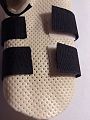
-
 Thenar eminence


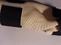
The strap locations are good for suspension and force application, although the wrist strap could be slightly more proximal. The thumb strap should be permanently attached on the medial aspect and the adhesive piece should be on the lateral side, making the donning and doffing of the device easier.
-
 Thumb strap permanent attachment

The client described the device as comfortable and good fit and did not report any discomfort to her styloid process of the radius which was within the trim lines.
Outcome measures
[edit | edit source]There are a variety of outcome measures available for the upperlimb functional ability. Choose an appropriate measure (if you are having trouble finding some the TAC website for clinical resources here may be useful.
The functional aims and goals of the client are to regain independence and perform daily activities and domestic chores. The Quadriplegia Index of Function (QIF) is comprised of 10 variables including grooming, bathing, feeding, dressing and personal care, which can be used as outcome measure for the client with a C7 injury. Grooming involved testing whether the patient was able to brush her teeth and brush her hair, bathing involved determining how well the patient was able to independently wash and dry her hair and body, feed herself and drink from a cup and assess her ability to take medications. Prior to receiving orthotic intervention to prevent contractures, the patient was unable to extend fingers enough to grasp larger objects, and lacked flexor power to grip. The low-temperature thermoplastic device was able to prevent contractures enabling the patient to extend fingers to hook her hand around objects, such as clothing for dressing and is able to bathe. However, for more precise tasks such as taking medication, feeding and grooming, she will need a definitive dynamic wrist-hand orthosis that will enable tenodesis of her hand to allow movement and grip. These improvements can be measured by assessing the patient’s ability to perform these tasks prior to treatment and following treatment.
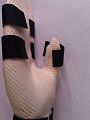
Below demonstrates the clients ability to extend fingers to grasp larger objects. The first image demonstrates little finger extension prior to orthotic intervention and the second image illustrates the effect of a resting splint. The patient's ability to actively move her fingers and wrist to a greater range can be accomplished with a dynamic wrist-hand orthosis.
-
 Limited finger extension
 -
 Finger extension and thumb abduction after resting splint

