
Upper Limb Orthotics/Extensor Carpi Ulnaris Tendonitis
Describe your case study
[edit | edit source]The patient being focused on throughout this case study is 38 year old Dennis. Dennis is a keen tennis player and is suffering from extensor carpi ulanaris (ECU) tendonitis due to the repetitive wrist motion used when hitting the ball. The client presented with ulnar sided wrist pain and is finding it difficult undertaking everyday tasks without experiencing significant pain. He has been advised by his doctor that a suitable brace must be fitted to ensure the wrist is immobilised. To provide the best opportunity for a successful recovery a brace must be fitted immediately, during the acute inflammation stage and worn for four weeks. In conjunction with bracing, the patient has been prescribed non-steroidal anti-inflammatory drugs. Dennis works as jeweller which requires significant fine motor skills of the hand. Therefore, opposition of the fingers must be considered when fabricating the orthosis.
Introduction
[edit | edit source]Extensor carpi ulnaris (ECU) tendonitis is found to be one of the most common sports related injuries directly caused by overuse (Parmelee-Peters & Eathorne, 2005; Wood & Dobyns, 1986). The injury presents itself with pain and swelling along the dorsal ulnar aspect of the wrist (Fulcher, Kiefhaber, & Stern, 1998, p. 441) and often affects people who participate in sports such as golf, tennis and rowing due to the repetitive wrist actions involved. For the client used throughout this case study, the mechanism of injury occurred whilst performing a double- handed backhand. Attempting to put spin on the ball meant the client’s dominant hand forcefully changed from a pronated position into supination. This sudden forearm rotation causes damage to the tendon (Campbell, Campbell, O’Connor, & Hawkes, 2013, p. 1107) Upon physical examination, ECU tendonitis will cause the person pain and discomfort whilst moving the wrist, especially during flexion and pronation (Wysocki, Biswas, & Bayne, 2012, p. 133). Clinical indications for the prescription of upper limb orthoses indicate 61% are due to traumatic injuries, with 39% of these being accounted for as tendon injuries such as this case (Bowker, Condie, Bader, & Pratt, 1993, p. 198).
Evidence
[edit | edit source]There are six tendon sheath compartments containing the tendons of the large extrinsic extensor muscles of the forearm, which cross the dorsum aspect of the wrist. The ECU is situated in the sixth dorsal compartment, proximally attaching on the lateral epicondyle of the humerus and inserting on the base of the fifth metacarpal (Reid, 1992, p. 1108). When orientating the ECU with landmarks, it is found on the medial border of the forearm, with its tendon running through the ulnar head and its styloid process (Moore, Dalley, and Agur, 2010, p. 756). The ECU muscle assists in wrist extension, acting with extensor carpi radialis brevis and longus to perform this movement. It also plays a role during ulnar deviation whilst providing stability to the ulnar side of the wrist.
ECU tendonitis as described by Fulcher et. al (1998) is the direct result of a microtrauma that occurs due to a stress or shear load. It often occurs when the tendon is overstretched during the deceleration phase of wrist flexion. Repetitive microtrauma to the tendon caused by collagen fibres sliding past each other causes inflammation, oedema and pain at the site thus resulting in tendonitis (Kannus, 1997, p. 78).
The pathogenesis process of ECU tendonitis can be divided into four different stages; inflammatory, proliferative, maturation and fibrosis. The inflammatory response is brought upon due to the microtrauma mentioned above (Retting, 2001, p. 592). For a successful recovery, treatment should be sought at this stage. Proliferation occurs in the following weeks, forming collagen in the affected areas. It is noted that activity must be limited at this stage. Following on from this, the maturation of collagen occurs, as it begins cross-linking and repairing tissue. At this stage of the process strengthening and stretching exercises should be performed to avoid muscle atrophy (Selvanetti, Cipollo, & Puddu, 1997, p.114).
Orthotic treatment options
[edit | edit source]In severe inflammatory cases, such as the client’s, Prentice (1994) outlines that it is crucial stressful forces such as chronic extension and overstretching are removed (p.359). For optimal healing of the tendon, the ulnar must be immobilised. An orthosis that aims to maximise radial hand function yet stabilise the ulnar is the ulnar gutter splint. Jacobs, Austin, and Austin (2003) describe the optimal positioning of the splint as 0-20˚ of wrist extension with slight ulnar deviation (p. 127). A more recent study conducted by Campbell et. al (2013) suggests bracing should hold the wrist in slightly more wrist extension of 30˚ (p. 1109). The design of this device ensures the medial aspect/ulnar side of the forearm is carefully stabilised, leaving the radial aspect of the device open. This device would be classified as a static orthosis, immobilising specific joints whilst allowing others to freely move (Jain, 1990, p. 259). This orthosis can be suitably made using low temperature thermoplastic such as the Orfit Ease (Orfit, 2011, para. 16). The rigidity of this material along with its ease of contouring provides the ideal support and immobilisation needed for this injury. Modifications are easily made to a device such as this, ensuring the best possible fit for the patient.
A prefabricated device such as the Ottobock Manu ComforT stable (Ottobock, 2014, para. 2) stabilises the wrist, restricting flexion and extension in a targeted method. This particular device covers the volar aspect of the forearm and hand, and is stabilised using a central strap. The Ossur Form Fit Wrist (Ossur, 2014, para. 5) works in a similar manner to support the wrist.
An inexpensive option in comparison to custom made and prefabricated orthoses is a Plaster of Paris (POP) cast. A short-arm plaster cast may be used to immobilise the arm in the same position used when working with thermoplastics. It has been proposed that a long-arm cast fitted to keep the forearm in a pronated position is more suitable as it allows the ECU to sit comfortably within the ulnar groove (Ankarath, 2006, p.150). Campbell et al. (2013) highlight that this theory is yet to be proven as there is no evidence suggesting the effectiveness of one method over another (p.1110).
Comparison of orthotic treatment options
[edit | edit source]Surgery is often required to treat more severe ECU injuries such as sublaxation due to the instability caused by the sub sheath tearing (Allende and Le Viet, 2005, p. 265). This treatment option however, is rarely required for ECU tendonitis as conservative management is proven to be successful on most occasions when implemented during the acute stage of inflammation (Fulcher et al., 1998, p. 441). In certain circumstances however, when the condition fails to respond to conservative management, leading to a chronic condition, Alexy and De Carlo (1998) recommend reconstructive surgery as the most suitable treatment option (p. 644).
Minimal research has been conducted concerning the effectiveness of custom made orthoses compared to pre-fabricated devices for a condition such as ECU tendonitis. Often times, prefabricated orthoses seem more appealing to client due to their sleek appearance and light weight however, a compromise must be made as they may not possess the specific features required to achieve a full recovery. A positive aspect of fabricating a custom made device such as the ulnar gutter design wrist-hand orthosis (WHO) is the variety of options on offer when choosing the most appropriate materials to use. For the given injury, rigid stabilisation of the ulnar would best be achieved using a high density polyethylene (HDPE) low-temperature thermoplastic. The materials used in the prefabricated orthoses described, provide stability to wrist but don’t possess the rigidity required for this given condition. The functionality of a POP cast is comparatively lower than that of the custom made WHO and prefabricated brace. Whilst it is recommended the devices are to be worn for the most part of the day, the ability to easily don and doff allows the patient to achieve everyday activities with ease. A POP cast in comparison, may significantly impede their ability to function. The client’s occupation as a jeweller requires him to perform fine motor skills, a custom made low temperature thermoplastic WHO with ulnar gutter will provide the stability needed to achieve a full recovery. The device will be customised to ensure full opposition of the fingers is achieved.
Search Strategy
[edit | edit source]A broad search was first conducted using libsearch, then refined using Medline, Cinahl and the Cochrane library. Key terms used were tendonitis/tendinitis,"extensor carpi ulnaris",ECU. ECU tendon, upper limb orthos*, splint and bracing.
References
[edit | edit source]Alexy, C., & De Carlo, M. (1998). Rehabilitation and Use of Protective Devices in Hand and Wrist Injuries. Clinics in Sports Medicine, 17(3), 635-655. doi: http://dx.doi.org/10.1016/S0278-5919(05)70106-X
Allende, C., & Le Viet, D. (2005). Extensor carpi ulnaris problems at the wrist–classification, surgical treatment and results. Journal of Hand Surgery (British and European Volume), 30(3), 265-272.
Ankarath, S. (2006). Chronic wrist pain: Diagnosis and management. Current Orthopaedics, 20, 141-151. doi:10.1016/j.cuor.2006.01.004
Bowker, P., Condie, D,N., Bader, D, L., & Pratt, D, J. (1993). Biomechanical Basis of Orthotic Management. Oxford, England: Butterworth, Heinemann.
Campbell, D., Campbell, R., O’Connor, P., & Hawkes, R. (2013). Sports-related extensor carpi ulnaris pathology: a review of functional anatomy, sports injury and management. Br J Sports Med, 47, 1105-1111. doi: 10.1136/bjsports-2013-092835
Fulcher, M, S., Kiefhaber, T. R., & Stern, P. J. (1998). Upper-Extremity Tendinitis and Overuse Syndromes in the Athlete. Clinics in Sports Medicine, 17(3), 433-448. doi: http://dx.doi.org/10.1016/S0278-5919(05)70095-8
Jacobs, M. A., Austin, N., & Austin, N. M. (Eds.). (2003). Splinting the hand and upper extremity: principles and process. Lippincott Williams & Wilkins.
Jain, A. S. (1990). Upper limb orthotics. Current Orthopaedics, 4(4), 259-262. doi: http://dx.doi.org/10.1016/0268-0890(90)90060-S
Kannus, P. (1997). Etiology and pathophysiology of chronic tendon disorders in sports. Scandinavian Journal of Medicine & Science in Sports, 7(2), 78-85. doi: 10.1111/j.1600-0838.1997.tb00123.x
Mintken, P. E., Glynn, P., & Cleland, J. A. (2009). Psychometric properties of the shortened disabilities of the Arm, Shoulder, and Hand Questionnaire (QuickDASH) and Numeric Pain Rating Scale in patients with shoulder pain. Journal of Shoulder and Elbow Surgery, 18(6), 920-926. doi: http://dx.doi.org/10.1016/j.jse.2008.12.015 Moore, K., Dalley, A., & Agur, A. (2010). Clinically oriented anatomy. Lippincott Williams & Wilkins.
Orfit. (2014). Physical Rehabilitation-Splinting Products. Retrieved from: http://www.orfit.com/en/physical-rehabilitation-products/
Ossur. (2014). Wrist and Thumb Support. Retrieved from: http://www.ossur.com/injury-solutions/products/upper-extremity/wrist-supports
Ottobock. (2014). Orthoses for joints, ligaments and fractures. Retrieved from: http://www.ottobock.com.au/cps/rde/xchg/ob_au_en/hs.xsl/38362.html
Parmelee-Peters, K., & Eathorne, S. W. (2005). The wrist: common injuries and management. Primary Care: Clinics in Office Practice, 32(1), 35-70.
Prentice, W, E. (1994). Rehabilitation Techniques in Sports Medicine. St. Louis, Missouri: Mosby.
Reid, D, C. (1992). Sports Injury Assessment and Rehabilitation. New York: Churchill Livingston
Rettig, A. C. (2001). Wrist and Hand Overuse Syndromes. Clinics in Sports Medicine, 20(3), 591-611. doi: http://dx.doi.org/10.1016/S0278-5919(05)70271-4
van Kampen, D.,A., Willems, W. J., van Beers, L.,W.A.H., Castelein, R. M., Scholtes, V. A. B., & Terwee, C. B. (2013). Determination and comparison of the smallest detectable change (SDC) and the minimal important change (MIC) of four-shoulder patient-reported outcome measures (PROMs). Journal of Orthopaedic Surgery and Research, 8, 40. doi:http://dx.doi.org/10.1186/1749-799X-8-40
Wood, M, B., & Dobyns, J, H. (1986). Sports-related Extraarticular Wrist Syndromes. Clinical orthopaedics and related research, 202, 93-102.
Wysocki, R. W., Biswas, D., & Bayne, C. O. (2012). Injection therapy in the management of musculoskeletal injuries: hand and wrist. Operative Techniques in Sports Medicine, 20(2), 132-141.
Functional Aims and Goals
[edit | edit source]During the initial stages of the injury, a plaster of paris (POP) cast may be fabricated as a temporary measure to stabilise the tendon. Throughout this stage, swelling is often an issue around the medial-dorsal aspect of the wrist, therefore securing the cast with material such as a non-compressive bandage will accommodate the inevitable fluctuation in volume. As a temporary device, the POP cast may indicate alterations that may be needed when making the definitive LTT wrist hand orthosis. Adjustments may include, high pressure areas requiring extra padding, areas restricting desired movements and inappropriate trimlines.
The main aim of the ulnar gutter LTT wrist hand orthosis (WHO) is to provide support to the ulnar/medial aspect of the wrist. As the WHO only provides stability over the medial aspect of the wrist an extra piece of LTT is added along the line of the ulnar to provide extra rigidity where the extra support is required. The distal trimline which is placed slightly proximal to the distal thenar crease allows for opposition of all the fingers as well as flexion at the metacarpophalangeal and interphalangeal joints. The patient is able to achieve optimal thumb function when placed in this position which maximises the patient’s ability to perform everyday activities. The proximal trimline is located about 5cm distally from the elbow, allowing a full range of motion at this joint.
Positioning the wrist in slight ulnar deviation aims to reduce the tension applied to the extensor carpi ulnaris, as the muscle is shortened in this position. Placing the wrist in this recommended position will ensure a faster healing time of the tendon. Movements such as wrist flexion and ulnar deviation are limited or even restricted when placed in this position as they have the ability to further aggravate the tendon. Whilst the device will stabilise and immobilise the ulnar to a certain degree, the aim is to also provide slight radial hand function. This is achieved through the design of the device which leaves the radial aspect of the wrist open.
Design
[edit | edit source]The trimlines of this device ensure a sleek appearance, as the client requires a simple and slim-line design. It important the distal trimline is located just proximal to the distal palmar crease as not to impede metacarpophalangeal movement. The trimline across the palm enables full thumb function, the thenar crease must be considered when marking this. The proximal trimline is located 2/3 the way down the forearm, this enables full elbow flexion and extension. The ‘ulnar gutter’ design covers the medial aspect of the wrist and forearm and only contacts half the circumference of the forearm. A permanent strap will be located through the web spacing between of the pollex and 2nd phalange. Removable padding made from ethylene-vinyl acetate (EVA ) will be used to cover this strap to reduce any rubbing that may occur. Removable straps are attached just proximal to the dorsal wrist crease and on the proximal trimline of the device. These straps will be made from Velcro loop. To ensure the ends of the straps don’t scratch the client, they will be rounded off. This will result in a more cosmetically pleasing appearance. Straps will be attached using a ‘sticky-back’ Velcro hook material.
Force Diagram
-
 Force System for orthotic device

- Numbered list item
Manufacturing process
[edit | edit source]•Comfortably position the patient and rest their hand on a flat surface. Trace their hand and forearm onto a piece of ‘Chux’ or a piece of paper. Ensure the tracing is done on the left side of the material, as extra material is needed for the template (Fig 1.)
•After tracing the hand and forearm, mark the proximal trimline, this is placed 2/3 the length of the forearm. Mark where the distal palmar crease lies, as the distal trimline falls just proximal to this crease. The thenar crease should also be marked, as this landmark guides the pattern of the design (Fig. 2)
•Once the patient removes their hand, the template of the orthosis can be transferred to the Chux. When drawing the template ensure the pattern is slightly wider on the proximal end as the forearm tapers distally. Add an extra 2cm to the length of the template as the distal trimline will be folded later in the process.
•Cut out the template and assess the fit, make adjustments accordingly.
•When satisfied with the template, trace the outline of it onto a piece of low temperature thermoplastic (LTT) (Fig 3) using a marker that can be easily removed. Cut the LTT and when ready to mould, place in a water bath set to approximately 60-70˚. (Fig 4)
-
 Figure 1
 -
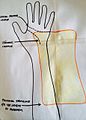 Figure 2
 -
 Figure 3


•Prepare your patient by positioning their hand in the desired position. Use a goniometer to ensure 20˚ wrist extension is achieved along with slight ulnar deviation of the wrist (Fig. 5). Place some padding over the ulnar styloid to provide a relief over this area. Remove a few layers to ensure it’s not too thick (Fig. 6).
•Carefully remove the warmed LTT from the waterbath, ensuring the material doesn’t adhere to itself and start draping the LTT over the ulnar border of the forearm and hand. Mould into the patient’s palm and check that the palmar material runs along the thenar line. Fold the distal trimline back so that it’s just proximal to the distal palmar crease.
-
 Figure 4
 -
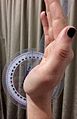 Figure 5
 -
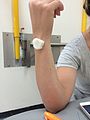 Figure 6


•Mark any excess material and remove the orthosis from the patient. Trim this material using a pair of sharp scissors.
•Reheat the proximal aspect of the orthosis by slightly dipping it into the water bath and gently flare the proximal edge.
•Cut a piece of thermoplastic that is measured to run the length of the orthosis, down the ulnar aspect. This piece will add extra rigidity and support down the medial aspect of the device (Fig 7). Heat the piece of LTT in the water bath then gently heat the area of attachment with a heat gun. When both are warm, mould the piece onto the orthosis.
•After leaving the orthosis to cool for a few minutes, fit to the patient once again, checking for correct alignment and comfort. Mark the location of the straps.
•Prepare 3 pieces of strapping, velcro loop for the web space, wrist and proximal edge of the orthosis. Using a heat gun, slightly warm the orthosis in areas where the Velcro hook is being attached. Avoid excessive heating, as this may deform the orthosis. Heat the sticky side of the Velcro hook and carefully place into position. For the strap located between the webspace, heat the loop side of the velcro and an area on the palmar aspect of the orthosis. Attach by pressing them together whilst they are still warm (Fig 8).
•Cut out a piece of low density EVA, and thread through the strapping of the webspace (Fig 9).
•Fit the orthosis to the client and assess any areas that may need to be adjusted.
-
 Figure 7
 -
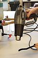 Figure 8
 -
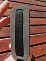 Figure 9


Finished Orthosis
-
 Coronal View - volar aspect
 -
 Sagittal View- Lateral aspect


Critique of fit
[edit | edit source]The client is a 38 year old male, who presented at the clinic with ulnar side wrist pain in his right hand (dominant hand). The client reports significant pain in his right wrist on the medial aspect. He reports that the injury is tennis related and that he has experienced pain in this area before. However, he believes it has gotten worse with time and now the pain is unbearable. He has stopped playing tennis and is finding it very difficult to perform his job as a jeweller as this requires fine motor skills and wrist actions. The patient would like to return to tennis, be able to perform his job more efficiently and reduce the pain he is currently experiencing.
A physical assessment was performed when the patient arrived, which revealed swelling, warmth and tenderness on the ulnar aspect of the wrist. An ECU synergy test was performed which requires the patient to flex their elbow at 90˚ whilst the examiner grasps the patients thumb and middle finger. The patient’s thumb is abducted against resistance. The client experienced pain along the dorsal ulnar aspect wrist when this was performed. Considering the clinical tests performed is it appropriate to conclude that the patient is indeed suffering from extensor carpi ulnaris tendonitis.
Due to the client’s line of work, I don’t want to fully restrict what he is able to do with his dominant right hand. Therefore, the goals will be to provide support to ulnar/medial aspect of the wrist, whilst allowing a bit of radial deviation to occur. The orthosis should allow full opposition of the digits so the client is able to perform the fine motor skills required as a jeweller.
Taking into account the orthotic goals of the client, an ulnar gutter wrist hand orthosis (WHO) seems appropriate. This design will restrict the movement of ulnar deviation whilst allowing the patient to achieve functional goals such as using all of his digits to perform fine motor skills such as adjusting and cleaning pieces of jewellery.
The custom made ulnar gutter WHO fits the prescription given however, there are a few changes that could improve this device. On the dorsal aspect of the WHO, the trimlines could have been extended more medially as seen in fig 1. Currently, the device slightly moves around in this area. Extending this trimline would help to reduce this movement. Due to the slim-line design of this device a thicker proximal strap would have been ideal to secure the device (fig 2). This material wasn’t available at the time but would have provided the extra support needed in this area.
-
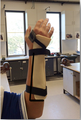 Figure 1
 -
 Figure 2


When fitting the device to the patient it became apparent that the strap between the webspace was causing some issues for the client as it felt uncomfortable. This was solved by attaching a piece of low density EVA to the strap. This addition appeared to fix the issue and the client was pleased with the outcome. The distal trimlines allow for full opposition of the fingers, which means the client can fulfill his work requirements (fig. 3). The extra rigidity down the ulnar side of the wrist was effective in stabilising the medial aspect of the wrist and reducing the movement of ulnar deviation.
-
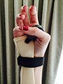 Figure 3
.JPG)
Outcome measures
[edit | edit source]Performing outcome measures on a client is an essential tool in determining treatment effects in research and the clinical setting by calculating a change in scores through pre and post treatment evaluation (van Kampen et al., 2009). For the subject in this case study it would be appropriate to use the quickDASH outcome measure before fitting the device and once the device is removed. This test aims to measure physical function and symptoms in people with upper limb disorders.
The patient undertook quickDASH test prior to having the orthosis fitted. At this stage the client was finding it difficult to perform everyday tasks and was experiencing significant pain in his wrist. His quickDASH disability/symptom score was 29.5 at this point. The test indicated that he was having difficulty performing tasks such as opening a jar, carrying shopping bags and experiencing mild-moderate pain in his wrist. Whilst this score is on the lower end of the scale, it indicated the client was having issues. A week after having his orthosis removed the test was repeated. The patient scored 6.8 which was a great outcome. He reported that he wasn’t experiencing any pain and wasn’t finding it difficult to perform activities that took some force or impact through the arm. According to a study performed by Mintken, Glynn, and Cleland, (2009) the minimum clinically important difference (MCID) for the quickDASH is a difference of 8 or more. Taking this into consideration, it is appropriate to assume the outcome is clinically important.
Before fitting the WHO, I performed a quick range of motion (ROM) test on patient’s wrist. In doing this, careful consideration was needed to ensure the patient didn’t injure himself further, and was instructed to stop when pain was experienced. The client only achieved 67˚ of wrist flexion before experiencing any pain, however full extension was achieved (70˚). The client struggled to achieve pronation, only reaching 75˚ along with 78˚ of supination. Full ROM was observed in the patient’s unaffected left wrist. During a follow up session, post the removal of the WHO, a vast improvement was observed in the client’s ROM of the right wrist. The patient didn’t report any pain whilst performing these tests and full ROM was achieved in both wrists.
Considering the results of the quickDASH and ROM tests performed before and after the intervention, it is appropriate to conclude that the client has made a full recovery.