
Upper Limb Orthotics/Dupuytren's disease
Describe your case study
[edit | edit source]The client is a 54 year old male basketball coach, of Scottish descent, who works at the local high school. He is fit and healthy in all aspects but has been suffering from the impact of Dupuytren's contractures for (approximately) the previous two (2) years. Dupuytren's is present in both hands, with the metacarpalphalangeal and proximal interphalangeal joints of the fourth and fifth digits on both hands being affected. The contracture's had not previously been to such a degree that they were overly impacting on the client's day to day activities, up until (approximately) 3 months ago, when he felt they were having a negative impact on his ability to coach basketball properly, and various other daily activities. The client expressed frustration about not being able to dribble or catch the ball properly, his driving was becoming increasingly difficult, and even the simple ability to put gloves on to keep himself warm was no longer possible. Upon measuring the angle of contracture at the MCP joints with a goniometer, it was found that his Dupuytren's had progressed to such a degree that surgery was warranted (Goniometer measurements were greater than the 30 degrees recommended for surgery). Upon successful completion of the surgery on both hands, it was suggested that orthotic management with a resting splint to maximise extension of the affected digits, would be beneficial to the client and aid him in returning to full functionality.
Written information
[edit | edit source]Dupuytren's Disease
[edit | edit source]Dupuytren’s disease or Dupuytren’s contracture is a progressive disease of the upper limb that affects the volar surface of the hands (Bayat & McGrouther, 2006; Warwick, Thomas & Bayat, 2012). It is a benign, generally painless, contractile condition of the palmar fascia that (Warwick et al., 2012; Worrell, 2012) results in permanent flexion deformity of the fingers (Bayat & McGrouther, 2006; Riolo, Young, Ueda & Pidgeon, 1991). There is no known cure for the disease and therefore much conjecture over the best form of management. The aetiology of Dupuytren’s disease is unkown (Evans, Dell & Fiolkowski, 2002), although it is apparent there is a genetic linkage (Bayat & McGrouther, 2006; Riolo et al., 1991; Swartz & Lalonde, 2008; Warwick et al., 2012), with a prevalence in white males of either a Celtic or Germanic ancestry (Bayat & McGrouther, 2006; Riolo et al., 1991; Swartz & Lalonde, 2008; Uzor, Maldjian, & Adam, 2011; Warwick et al., 2012; Worrell, 2012). Age is also a determinant factor, with there being a higher prevalence in individuals after the age of fifty (Riolo et al., 1991; Swartz & Lalonde, 2008; Uzor et al., 2011; Warwick et al., 2012; Worrell, 2012). There are associated risks connected with Dupuytren’s contractures to diseases such as epilepsy, diabetes, alcoholism and smoking (Riolo, et al., 1991; Uzor et al., 2011; Warwick et al., 2012) although the exact connection is still not completely understood.
Pathophysiology
[edit | edit source]Various studies indicate that the pathophysiology involved in Dupuytren’s disease is a similar process that occurs with normal wound healing (Bayat & McGrouther, 2006; Warwick et al., 2012), - an increased proliferation of fibroblasts; which then differentiate into myofibroblasts; causing collagen deposition which results in the contractural cord like structures (Bayat & McGrouther, 2006; Riolo et al., 1991; Warwick et al., 2012).
Clinical Presentation
[edit | edit source]The clinical presentation of Dupuytren’s disease starts with the appearance of nodules on the palm, which is a result of the palmer fascia thickening (Bayat & McGrouther, 2006; Swartz & Lalonde, 2008;), usually at the base of either the small, or ring finger (Riolo et al., 1991). Further thickening of the palmer fascia leads to formation of palpable cord like structures, which extend into the affected fingers from the palm (Bayat & McGrouther, 2006; Riolo et al., 1991; Swartz & Lalonde, 2008), and can cause contractures of the metacarpophalangeal joints and/or the proximal interphalangeal joints (Riolo et al., 1991; Swartz & Lalonde, 2008; Uzor et al., 2011; Warwick et al., 2012), with the fingers most commonly affected being the fourth (ring finger) and fifth (little finger) digits (Bayat & McGrouther, 2006; Swartz & Lalonde, 2008; Worrell, 2012). Dupuytren’s disease can affect either or both hands, with over 50% of people presenting with a bilateral condition, (Bayat & McGrouther, 2006; Riolo et al., 1991) and contractures can progress until the fingertips will ultimately touch the palm (Riolo et al., 1991). As is the case with this patient, most people with Dupuytren’s disease generally do not seek assistance until the contracture begins to impact on their daily lives, making simple tasks like washing their face, combing their hair or driving, harder to perform (Ball, Pratt & Nanchahal, 2013; Bayat & McGrouther, 2006; Swartz & Lalonde, 2008).
Testing & Measuring
[edit | edit source]Simple tests and measures that can be performed to indicate the presence of Dupuytren’s disease vary between clinicians but can include the Hueston Table Top Test (Bayat & McGrouther, 2006; Hueston, 1976 as cited in Brenner, 2002; Swartz & Lalonde, 2008; Warwick et al., 2012;), whereby a patient endeavors to place their palm flat on a table, and if unable to do so the test is positive. Range of motion measurements using a goniometer to measure the affected joint angles, is also an effective means to note the disease progression (Brenner, 2002; Warwick et al., 2012), which can lead to further classification by Tubiana (Tubiana, 1986 as cited in Warwick et al., 2012) of the four categories of deformity – Stage I: 0-45°; Stage II: 45-90°; Stage III: 90-135° and Stage IV: 135-180° (Tubiana, 1986 as cited in Warwick et al., 2012).
Treatment Options
[edit | edit source]There is no known cure for Dupuytren’s disease but it can be managed, to varying degrees of success, with a variety of clinical alternatives (Bayat & McGrouther, 2006; Larocerie-Salgado & Davidson, 2011; Riolo et al., 1991; Swartz & Lalonde, 2008; Warwick et al., 2012), the most basic of which is to simply observe and monitor the condition of the hand, if only nodules are present and functionally impairing contractures are not yet apparent (Swartz & Lalonde, 2008; Warwick et al., 2012). According to Larocerie-Salgado and Davidson (2011), the use of night time extension splints in conjunction with stretching and massage of the contracted tissue, can delay disease progression and improve mobility for individuals in the early stages of Dupuytren’s disease, though most other reviewed literature indicates that orthotic management only comes into play postoperatively (Bayat & McGrouther, 2006; Clare, Hazari & Belcher, 2004; Swartz & Lalonde, 2008; Worrell, 2012). Other clinical management options, which are gaining wider understanding and acceptance, include radiotherapy - which is thought to be only effective in early stages of the disease before contracture occurs; steroid injection – again only thought to be effective in early disease stages; and injectable collagenase – which directly dissolves the abnormal collagen within the thick cord like structures (Warwick et al., 2012). All of these options require further testing to provide any conclusive basis for evidential practice. The gold standard for management of Dupuytren’s disease is that of surgery, (Bayat & McGrouther, 2006; Swartz & Lalonde, 2008) with metacarpophalangeal contractures of 30° and above being generally indicative that surgery would be of benefit (Riolo et al., 1991; Swartz & Lalonde, 2008). Surgery tends to gain functional improvement for the patient, which includes achieving not only extension of the affected digits, but also maintaining some degree of flexion so that the hand is a useful and functioning part of the body (Bayat & McGrouther, 2006). Surgical options include divisions of the affected fascial cord (fasciotomy), excisions of the diseased fascial cord (partial or total fasciectomy), or excision of both the diseased fascial cord as well as the overlying skin (dermofasciectomy) (Bayat & McGrouther, 2006; Jerosch-Herold et al., 2011; Swartz & Lalonde, 2008; Warwick et al., 2012). Although there is a great degree of success with any of the operative measures for releasing Dupuytren’s contracture (Bayat & McGrouther, 2006; Swartz & Lalonde, 2008; Warwick et al., 2012), recurrence of the disease is relatively high (Bayat & McGrouther, 2006; Swartz & Lalonde, 2008), which can ultimately lead to amputations (Swartz & Lalonde, 2008).
Orthotic Management
[edit | edit source]According to the literature it is postoperatively, that orthoses are used in the management of Dupuytren’s disease (Bayat & McGrouther, 2006; Clare et al., 2004; Swartz & Lalonde, 2008; Worrell, 2012). The use of thermoplastic extension splints at night and/or daytime, as well as regular therapy sessions with a hand therapist/physiotherapist, may aid in the recovery process postoperatively (Bayat & McGrouther, 2006; Jerosch-Herold et al., 2011), although there is much conjecture within the reviewed literature as to whether there is any greater benefit to splinting than purely hand therapy alone (Jerosch-Herold et al., 2011; Kemler, Houpt & van der Horst, 2012; Larson & Jerosch-Herold, 2008; Warwick et al., 2012). Within the literature there is no general consensus on the type of orthoses to be used nor the duration of use (Kemler et al., 2012; Larson & Jerosch-Herold, 2008), though it would seem apparent that an orthosis designed to maximise extension of both the metacarpophalangeal and proximal interphalangeal joints would be appropriate (Clare et al., 2004; Jerosch-Herold et al., 2011).
Comparison of Orthotic vs Surgical Management
[edit | edit source]In comparing the orthotic and surgical management, there is still much conjecture over the best means to achieve the greatest functional outcome for those presenting with Dupuytren’s contracture, or whether indeed orthotic intervention has any place within the management protocol. Currently surgery is the standard when it comes to providing management, though much research is still needed to come to any conclusive decisions about orthotic management of Dupuytren’s disease.
References
[edit | edit source]Ball, C., Pratt, A., & Nanchahal, J. (2013). Optimal functional outcome measures for assessing treatment for Dupuytren’s disease: a systematic review and recommendations for future practice, BMC Musculoskeletal Disorders, 14, pp1-11. doi: 10.1186/1471-2474-14-131
Bayat, A., & McGrouther, D. (2006). Management of Dupuytren’s disease – clear advice for an elusive condition, Annals of the Royal College of Surgeons of England, 88, pp3-8. doi: 10.1308/003588406X83104
Brenner, P. (2002). Dupuytren’s disease of ring and little finger, Orthopedics and Traumatology, 2, pp138-158. Retrieved from
Clare, T., Hazari, A., & Belcher, H. (2004). Post-operative splinting to maintain full extension of the PIPJ after fasciectomy, British Journal of Plastic Surgery, 57(2), pp179-180. doi: 10.1016/j.bjps.2003.11.013
Evans, R., Dell, P., & Fiolkowski, P. (2002). A clinical report of the effect on functional results after fasciectomy for Dupuytren’s contracture, Journal of Hand Therapy, 15(4), pp331-339. Retrieved from http://0-search.proquest.com.alpha2.latrobe.edu.au/docview/222230445?accountid=12001
Jerosch-Herold, C., Shepstone, L., Chojnowski, A., Larson, D., Barrett, E., & Vaughan, S. (2011). Night-time splinting after fasciectomy or dermofasciectomy for Dupuytren’s contracture: a pragmatic, multi-centre, randomised controlled trial, BMC Musculoskeletal Disorders, 12 (136), pp1-9. doi: 10.1186/1471-2474-12-136
Kemler, M., Houpt, P., & van der Horst, C. (2012). A pilot study assessing the effectiveness of postoperative splinting after limited fasciectomy for Dupuytren’s disease, Journal of Hand Surgery (European Volume), 37(8), pp733-737. doi: 10.1177/1753193412437631
Larocerie-Salgado, J., & Davidson, J. (2011). Nonoperative treatment of PIPJ flexion contractures associated with Dupuytren’s disease, The Journal of Hand Surgery (European Volume), 37E(8), pp722-727. doi: 10.1177/1753193411422680
Larson, D., & Jerosch-Herold, C. (2008). Clinical effectiveness of post-operative splinting after surgical release of Dupuytren’s contracture: a systematic review, BMC Musculoskeletal Disorders, 9, pp104-108. doi: 10.1186/1471-2474-9-104
Riolo, J., Young, L., Ueda, K., & Pidgeon, L. (1991). Dupuytren’s Contracture. Southern Medical Journal, 84 (8), pp983-996. Retrieved from: http://0-ovidsp.tx.ovid.com.alpha2.latrobe.edu.au/sp-3.11.0a/ovidweb.cgi?WebLinkFrameset=1&S=FHLJFPNDFBDDBNFJNCMKHFGCDMILAA00&returnUrl=ovidweb.cgi%3fMain%2bSearch%2bPage%3d1%26S%3dFHLJFPNDFBDDBNFJNCMKHFGCDMILAA00&directlink=http%3a%2f%2fgraphics.tx.ovid.com%2fovftpdfs%2fFPDDNCGCHFFJFB00%2ffs047%2fovft%2flive%2fgv038%2f00007611%2f00007611-199108000-00011.pdf&filename=Dupuytren%27s+Contracture.&navigation_links=NavLinks.S.sh.22.1&link_from=S.sh.22%7c1&pdf_key=FPDDNCGCHFFJFB00&pdf_index=/fs047/ovft/live/gv038/00007611/00007611-199108000-00011&D=ovft&link_set=S.sh.22%7C1%7Csl_10%7CresultSet%7CS.sh.22.23%7C0
Swartz, W., & Lalonde, D. (2008). Dupuytren’s Disease, Plastic and Reconstructive Surgery, 121 (4), pp1-10. doi: 10.1097/01.prs.00003405932.46121.84
Uzor, R., Maldjian, C., & Adam, R. (2011). Imaging for Dupuytren contracture, The Journal of Muscoloskeletal Medicine, 28(2), pp56-57. Retrieved from http://0-search.proquest.com.alpha2.latrobe.edu.au/docview/863650370
Warwick, D., Thomas, A., & Bayat, A. (2012). Dupuytren’s disease: overview of a common connective tissue disease with a focus on emerging treatment options, International Journal of Clinical Rheumatology, 7(3), pp309-323. doi: 10.2217/ijr.12.25
Worrel, M. (2012). Dupuytren’s Disease, Orthopedics (online), 35 (1), pp52-60. Retrieved from http://0-search.proquest.com.alpha2.latrobe.edu.au/docview/914685756
Search Strategy
[edit | edit source]A literature search was conducted of the Medline and Pubmed databases along with Google Scholar, using the search terms of Dupuytren's Disease, Dupuytren's Contracture, Orthoses, Orthotic Devices, Splints, Hand Therapy and Orthotic management. Appraisal of the acquired literature was conducted, and the references perused to ascertain if any other relevant literature was missed in the initial search.
Functional Aims and Goals
[edit | edit source]The functional aim for the production of the hand based Dupuytren’s orthosis is to maintain the range of motion that has been achieved post surgery for both the metacarpophalangeal joints and proximal interphalangeal joints of both the fourth and fifth digits of both hands of the client. The goal of surgery was to release the flexion contractures that were present at both the metacarpophalangeal and proximal interphalangeal joints, this was achieved, therefore maintenance of the joints in as great a degree of extension as possible, to prevent contracture reformation, is the aim of the orthosis. The use of the hand based orthosis at nighttime, and as required during the day, will prevent the fingers from flexing and hence the promotion of extension of the digits. Whilst, for this case study, the orthosis was manufactured out of both plaster of paris and a low temperature thermoplastic, the material of choice for the long term manufacture/maintenance of Dupuytren’s contracture, would be that of the low temperature thermoplastic due to the material being lightweight, the subsequent ease of donning/doffing and the ease of maintenance of the orthoses.
Design
[edit | edit source]Whilst both hands of the client are affect by Duputreyn's contracture, due to time constraints, only the design and manufacturing process for the left hand are being discussed - only a left hand orthosis was manufactured.
Design:
The design prescription for the device was for a custom made hand orthosis manufactured from low temperature thermoplastic. It was designed with the aim of allowing the 4th and 5th digits of the left hand to rest in maximal extension, at both the metatarsal phalangeal joints and the proximal interphalangeal joints, to aid in maintaining the contracture release that was achieved from surgery.
Trimlines:
The trimlines of the device are done to maximise comfort for the client, allow the fourth and fifth digits to rest comfortably, and not to impede on any movement at the wrist joint or thumb.
The proximal trimline of the device should sit distal to the wrist joint complex, allowing the normal transverse arch of the palm to occur.
The distal trimlines are the distal palmar crease extending to fully encompass the length of the fourth and fifth digits, care being taken as to not encroach on the middle or third digit, and allow its full range of motion to still occur.
Velcro straps are required for secure attachment of the device, with a permanent strap to be secured on the dorsal aspect of the device, and an adjustable, removable strap to be secured around the digits.

Force Systems. 
Original design volar aspect 
Original design dorsal aspect


Manufacturing process
[edit | edit source]Materials required:
- Chair & table (for comfort of patient), pen, kitchen “chux” for pattern, scissors, suitable size of lower temperature thermoplastic (LTT), electric frypan with suitably heated water, Velcro, heat gun
- With patient seated comfortably, have them rest their hand on a suitable size of the “chux”, their fingers resting comfortably and their thumb extended as far as possible (Figure 1)
- Trace around the hand, marking the wrist joint, the distal palmar crease, the metatarsalphalangeal joints & the interphanlangeal joints of the affected digits (in this case the fourth and fifth), and the metatarsalphalangeal joint of the thumb (Figure 2 & 3)
- On the chux, draw a pattern encompassing the entire palmar aspect of the fourth and fifth digits, down to the distal palmar crease
- Extend the line of the distal palmar crease out to the web space between the thumb and second digit
- Make a tab that extends out from this, that will sit in the web space and wrap around to the dorsal aspect of the hand
- The trimline for the proximal edge of the device extends from the MTP joint of the thumb across to a point approximately 1cm distal to the wrist joint, again with another smaller tab extending out from this
- Cut the pattern out and check that it suitably fits the patient. Adjust trimlines if necessary.
- When the pattern is correct, trace around it onto the piece of low temperature thermoplastic and then cut to size with scissors (Figure 4)
- Place in the heated water to soften
- Whilst this is softening, get the patient to sit with their elbow resting comfortably on either the armrest of the chair (if it has one), or on the edge of the table, and place their hand into a supinated position with the affected digits as maximally extended as possible
- Take the LTT out of the water, and dry quickly, and proceed to fold the edges of the LTT over so as to prevent the edges rubbing and annoying the patient. This needs to be done on the entire length of the proximal trimline, and the distal trimline that extends across the distal palmar crease and up the lateral edge of the fourth finger. This needs to be done promptly.
- Once the edges are suitably folded, place the warm LTT on the patients hand, align the piece correctly, and then proceed to carefully mould the LTT around the medial and lateral sides of the hand and hold securely in place whilst waiting for the LTT to cool (Figure 5, 6 & 7)
- Check for comfort and ensure nothing is causing pressure or rubbing, take the device off the patient
- Cut a suitable length of LTT to reinforce the strength of the finger section and permanently adhere using a heat gun to gently heat both sides required for attachment (the orthosis and the extra LTT piece)
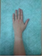
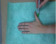
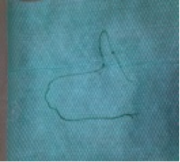
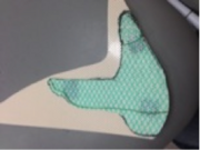


Fig 1 Fig 2 Fig 3 Fig 4 Fig 5 Fig 6 Fig 7


Finishing process:
- Once the orthosis has cooled, be sure that achieves the function it needs to and then mark out where the placement of the straps needs to be
- Cut the Velcro straps to suitable size
- Using a heat gun, warm the medial aspect of the hand tab on the LTT, and the piece of Velcro, and attach together to form a permanent bond
- Place the Velcro hook on the thumb tab by using the same technique with the heat gun
- The Velcro hook for the finger strapping needs to be placed on the volar aspect of the reinforce fingers, at a suitable height for the patient that it doesn’t rub and aggravate the web space between the 3rd and 4th digits
- Cut a suitable length of Velcro that can be attached around the finger, comfortably encompassing both fingers, that will adhere to the hook from either side
- Clean any pen marks off the device and do a final check on the patient, ensuring the device is comfortable and easy for them to don and doff

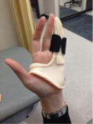
Volar view of finished orthosis 
Dorsal view 
Volar view 
Saggital view


Critique of fit
[edit | edit source]The client is a 54 year old male who recently had surgery (fasciectomy) to regain functional improvement from the effects of Dupuytren’s contracture at both the MCP & PIP joints, bilaterally. Surgery was successful, with full ROM restored to all joints and fingers, and the client has been referred onto us for orthotic management to help maintain this ROM, and prevent joint flexion contractures from reoccurring.
Upon presentation to the clinic, the patient was optimistic about his future being free from the contractures associated with Dupuytren’s and was keen to utilise orthotic management to help aid in this process. Observation and measurements taken at the affected joints indicated that ROM at both MCP & PIP joints was normal, and wound healing from the operation was complete.
The goal of the orthotic management is to maintain the MCP & PIP joints, on both hands, in as much extension as possible, to help prevent the flexion contractures associated with Dupuytren’s disease from impacting on the daily activities of the patient (e.g. Coaching basketball, driving and putting on gloves).
In order to achieve these goals, an orthotic prescription has been developed, to provide resting orthoses for the client. As there is much conjecture within the literature as to the benefits of orthotic management of Dupuytren’s disease, and therefore no design principles to go off, a prototype design was manufactured that aimed to encompass the needs of maintaining the affected joints at maximum extension, to be worn by the client predominately as a night time resting orthosis, or as required during the day if the client felt the need. It was manufactured out of low temperature thermoplastic.
The custom made hand orthosis fits the orthotic prescription, though there are a few areas in which the device could be improved. As it was a prototype device, with no precursor to work from, some measure of trial and error is to be expected.
The anterior proximal trim line is adequate, and does not impinge on any movement at the wrist. Where it anteriorly crosses the proximal edge of the web space between the thumb and index finger, it restricts movement somewhat. The restriction is only minimal as the thumb can still be utilised in holding and gripping things, but the trimline, as it sits, would be potentially uncomfortable after a period of time.

Area where slight impingement occurs over web space

Posteriorly the proximal trimline, again is adequate, as no movements are impinged upon, but the lateral strap attachment, pushes into the dorsal surface of the hand and causes unnecessary pressure, which again over time would be considerably uncomfortable for the client.

Area where dorsal "pushing" is evident

The distal trimlines are good, sitting proximal to the distal palmer crease and extending further to encompass the fourth and fifth phalanges. The fingers sit comfortably and the lateral edge of the thermoplastic has been rolled so that there is no rubbing or irritation to the middle finger. The strapping is in a comfortable position and does not rub on the web space, though care must be taken to ensure this does not happen in future design.
Potentially one drawback of the design is in only immobilising the affected ring and little fingers – as the middle finger shares a tendon with the ring finger, the ring finger tends to want to move when the middle finger does, but as it is restricted it can’t. There is some concern as to whether this might cause strain on either of the two fingers and whether or not the design should immobilise the middle finger as well.
The technical work on the LTT orthosis is acceptable. The edges are finished off well, there are no points for irritation, other than those discussed above, and the straps securely attach the device to the client.
The hand orthosis achieves its function – the client is unable to flex the MCP & PIP joints and these joints are maintained in maximum extension.
The client states that the device is comfortable to wear, apart from the minor issues as discussed above. The design is a prototype and with insufficient design guidelines available from the literature, it seems a fair attempt at providing appropriate immobilisation as required for this Dupuytren’s client.
Volar view of finished orthosis 
Dorsal view of finished orthosis


Outcome measures
[edit | edit source]Due to there being much conjecture in the literature as to whether there is any clinical benefit to the use of hand orthoses for people with Dupuytren’s disease, it is difficult to effectively assess what outcome measures should be used for this client. The symptoms the client was presenting with were prior to surgery for the contractures and whilst we have grading we can make comparisons on, they are probably more directly rated to the effects of surgery, rather than orthotic intervention. Nevertheless, such outcome measures will be discussed below.
Pre surgery: The outcome measure the client was assessed with, prior to surgery, was the Disabilites of the Arm, Shoulder and Hand Questionnaire (DASH) – it is considered a valid and reliable tool to assess improvements (or not) in functions for those with upper limb impairments. The higher a score is on the DASH, the greater the disability.
Prior to surgery the client scored a 50.8 on the questionnaire, indicating a moderate-severe level of impact on their daily life; with higher scores on the optional work & sports modules of the DASH – being 68.8 and 81.3 respectively.
As well as the DASH, the clients accounts of his frustrations about being able to not function in his job and life were taken into account, and the main “outcome measure” for the client was in being able to get full motion back into his hands to enable him to perform both work and daily activities with functional normality.
Post surgery: As indicated earlier, surgery for the clients’ contractures was successful, and was achieved in giving back full ROM at the MTP and PIP joints. After successful healing of the wounds from surgery, the client again undertook the DASH and the resultant scores were 0.8, 0 & 0 – which are obviously a marked improvement on the original scores. A change of greater than 15 in the DASH is considered to signify a clinically important difference, and in this case, surgery could be considered a great success.
Orthotic intervention: The client presented to us in the hopes of preventing contracture reformation, and as detailed, hand orthoses were constructed. They were provided to the client mainly as a “resting” orthoses, with the outcome of the patient having the ability to don them of a nighttime, or as he felt he needed during the day, to aid in the maintenance of the extension achieved by surgery of the affected digits and joints. According to the client they are “working beautifully”, and definitely keep the fingers extended to their maximum capacity. He considers them of benefit in his rehabilitation, and is using them consistently in conjunction with physiotherapy work being done by a hand therapist. The outcome measures and aim, appear to have been achieved for this client.
Referral Letter
[edit | edit source]27/5/14
Jen Smith
Physioworks
Sladen Street, Northcote
Dear Jen,
I am referring onto you my client, a 54 year old male who has recently had fasciectomy surgery (3/12) to release the flexion contractures present at the MCP joints and the PIP joints at the fourth (4th) and fifth (5th) digits bilaterally. The flexion contractures were caused by Dupuytren’s disease. Surgery was successfully performed at Box Hill Hospital (3/12) with the flexion contractures being released and full ROM being achieved at all affected joints. On presentation to our clinic, the client expressed his willingness to work with necessary allied health professional in maintaining this normalised ROM and prevent the flexion contractures from returning. Custom made LTT “resting” hand orthoses were fabricated – a prototype designed with the aim of maintaining full ROM and thus maximum achievable extension of the affected joints. The client has been advised to wear the orthoses at night time and as needed during the day. As an adjunct to orthotic management, I believe it is in the best interest of the client, to work with you receiving physiotherapy, as you see fit, to maintain this full ROM that surgery achieved. If, however, you notice that the clients recovery is detioriating or not progressing as expected, I ask that you refer back to the consulting surgeon at Box Hill Hospital (Dr Took), to ensure there are no complications from surgery. If you have any questions regarding the management of this client, please don’t hesitate to contact me.
Kind regards,
Nicole Reynolds