Electrical impedance tomography
Electrical Impedance Tomography
[edit | edit source]Introduction
[edit | edit source]Electrical Impedance Tomography (EIT) or Bioimpedance Tomography is an imaging method that aims to reconstruct the electrical conductivity distribution of an object based on electrical voltage measurements made on its surface when an electrical current is applied to it [1]. It has many fields of application including industry, geophysics and medicine [2]. Although it is not yet used in everyday clinical practice, it is considered to have a big potential as it is a relatively low-cost and minimally invasive technique [1].
Forward Problem.
[edit | edit source]The Forward Problem (FP) consists in predicting the electrical voltage distribution on the outermost surface of the object when the conductivity distribution and the applied current are known. Maxwell’s equations govern the physics of the problem.

If the object has a special geometry such as spherical or cylindrical, the FP can be solved analytically [3] [4] [5]. However, the objects under study (e.g. thorax or brain) are not usually found in these convenient shapes, although it is common practice to approximate them as spheres or cylinders. If the real shape is going to be considered, 2-D or 3-D electrical models of the object must be built and numerical methods such as the Finite Element Method (FEM) or the Boundary Element Method (BEM) can be used to solve the FP over arbitrary geometries. [6][7][8]
Inverse Problem.
[edit | edit source]The Inverse Problem (IP) or reconstruction problem consists in the estimation of the internal conductivity based on measurements of the electrical voltage on the surface and the knowledge of the applied current. The general idea is to find the conductivity that minimizes the difference between the measured and predicted (by the FP) voltages [2]. As some other inverse problems, it is ill-conditioned so that some a priori information is needed to convert it to a well posed problem. Several algorithms have been proposed and discussed in the literature [2]. The IP can be concerned with the estimation of the entire conductivity distribution (or conductivity map) or with the estimation of a few key parameters. This parametric approach comprises, for example, the estimation of coefficients of smooth basis functions of conductivity, or the estimation of the conductivities of the main tissues of the head when the assumption of constant conductivity within each tissue is made [6][8].
Application to brain function.
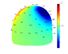
[edit | edit source]When EIT is applied to the study of brain function, the most common set-up consists in several electrodes attached to the scalp connected to a current generator and a data acquisition system. At a time , two electrodes drive the current while the electrical voltage is measured on the others. In zones of the brain where increasing activity occurs, there is an increment of blood that produces a slight change in the impedance of that region. Although difficult to measure, this impedance change could be detected by EIT [1][9]. Small and fast conductivity changes are also produced by neuron depolarization and at present, EIT is the only method that has the potential to image them [9]. The parametric approach can be used to estimate the conductivity of the main tissues of the head, based on the knowledge of the surfaces that delimit them. This information can be obtained, for example, from Magnetic Resonance Images.
References
[edit | edit source]- ↑ 1.0 1.1 1.2 Bayford, R. Bioimpedance tomography (electrical impedance tomography). Annu. Rev. Biomed. Eng., 8:63–91 (2006).
- ↑ 2.0 2.1 2.2 Lionheart WRB, Polydordes N, Borsic A. The reconstruction problem. Electrical Impedance Tomography: Methods, History and Applications, ed. DS Holder, Part 1, Inst. Phys., pp. 3–64 (2004).
- ↑ E. Frank. Electric potential produced by two point current sources in a homogeneous conducting sphere. J. Appl. Phys., 23(11):1225-1228 (1952).
- ↑ J.C. de Munck, T.J.C. Faes, R. Heethaar. The boundary element method in the forward and inverse problem of electrical impedance tomography. IEEE Trans. Biomed. Eng., 47:792–800 (2000).
- ↑ Fernández-Corazza M, von Ellenrieder N, Muravchik CH. Estimation of electrical conductivity of a layered spherical head model using electrical impedance tomography. J Phys Conf Ser, 332(1):012022 (2011).
- ↑ 6.0 6.1 S.I.Gonçalves, J.C. de Munck, J. Verbunt, F. Bijma, R. Heethaar, F. L. da Silva. In vivo measurement of the brain and skul resistivities using an EIT-based method and realistic models for the head. IEEE Trans. Biomed. Eng., 50(6):754–767 (2003).
- ↑ Abascal JFP, Arridge SR, Atkinson D, Horesh R, Fabrizi L, Lucia MD, et al. Use of anisotropic modelling in electrical impedance tomography; description of method and preliminary assessment of utility in imaging brain function in the adult human head. NeuroImage, 43(2):258 –68 (2008).
- ↑ 8.0 8.1 Fernández-Corazza, M., L. Beltrachini, N. von Ellenrieder, y C. H. Muravchik. Tomografía de impedancia eléctrica y resonancia magnética como herramientas conjuntas para la estimación paramétrica de la conductividad eléctrica del cráneo y del cuero cabelludo. In XIV Reunión de Trabajo Procesamiento de la Información y Control RPIC 2011, tomo 845–850, págs. 1–6. Oro Verde (2011).
- ↑ 9.0 9.1 Holder, D. Electrical impedance tomography of brain function. In World Automation Congress. Hawaii (2008).
