Cell biology/Membrane Transport: Permeases and Channels
Here is the link to the ITunes U Lecture from Berkeley. Membrane Transport: Permeases and Channels
Sisterhood of the Traveling Proteins Part II
[edit | edit source]Experiment from last lecture reviewed.
- It is possible to make a Heterokaryon cell with mouse and human cells. The human cell proteins can be visualized with antigens that are attached to a dye, FlTC which fluoresces yellow-green. Mouse cell proteins can also be marked with antigens tagged with rhodamine which fluoresces red.
- Sendai virus can cause cells to fuse via a cell junction (if there are a lot of cells around-the virus actually trying to fuse to a cell surface to inject the RNA core).
- The rate of mixing of antigens (attached to proteins in the cellular membrane) is roughly determined by viscosity of the phospholipids. If you cool the proteins with ice, the proteins don't redistribute in the cell membrane. If you add an energy poison that blocks the motile apparatus of the cytoskeleton, the proteins in the cellular membrane still mix.
- This study shows that proteins are very fluid within the cellular membrane and don't require ATP consumption.
- This experiment is not very precise because you create antibodies for a variety of different proteins.
- However this technique can be used with more specificity if you choose a single protein to monitor.
- This technique is very similar to the experiment above but can be monitored with a very specific rate. A laser is shined onto the cell membrane frying out the flourescent molecules but not damaging the cellular membrane. Immediately there is a dark area in the membrane but over time the dark spot goes away because the fluorescing and nonfluorescing proteins mix.
- This technique allows a very precise measurement of the diffusion of proteins within the cellular membrane, and it works with most cellular proteins.
- Exception
- The Band 3 anion transport channel is tied to the spectrin cytoskeleton network. This protein is anchored in place and if you destroy the fluorescing antigen other antigens won't flow into that location.
Fluid Mosaic Model of Membrane Structure
[edit | edit source]
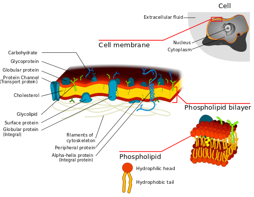
- Proteins are able to diffuse throughout the cell through two dimensions of movement (cannot reverse inner-outer orientation). Sometimes proteins are held in place by a cytoskeleton and then they are not able to flow as freely throughout the membrane.
Corrections to this model
- Cholesterol and Sphingolipids form a distinct phase in the bilayer called "rafts" and they float throughout the membrane and some proteins have an affinity for these "glycolipid rafts" and they float around the membrane within these spots and are not free to interact with the phospholipids in the rest of the membrane. These rafts are likely to have important functions like improving cell signaling but it is an area still under research.
Transport
[edit | edit source]Passive
[edit | edit source]Pores
[edit | edit source]Pores (such as porin) allows small molecules into/out of the cell and are not selective. Pores will allow molecules to be transferred in and out of the cell until equilibrium is reached.
Facilitated Carriers
[edit | edit source]A protein recognizes a particular small molecule (like glucose) and this protein allows molecule to flow into cell until equilibrium reached inside and outside the cell. No energy is consumed and the process is selective.
Active
[edit | edit source]Transport is concentrating a molecule against it's concentration gradient and is an energy driven process.
Direct
[edit | edit source]Direct Active Transport is mainly ATP-driven permeases
- F type (and V type) this type is like the type in mitochondria that pump protons into the intermembrane space and are able to form ATP molecules when the protons flow in through that channel. The V type pumps protons into a vacuole to cause it to become acidic and is very similar to the F-type. Both are formed by large complexes of proteins.
- P type- ATP is hydrolyzed and covalently attached to an amino acid, aspartamine, which activates a protein and channel changes shape causing it to start functioning
- ABC type- ATP binding cassette-this can help create channels to expel toxins. It creates a problem with chemotherapy because the chemotherapy agents are forced out of the cell. It has an ATP binding site that directly drives this protein.
Linked to a Gradient
[edit | edit source]Facilitated carrier becomes active by linking the uptake of a desired molecule with another that has a large concentration difference across the membrane (see below).

- It is a P-type permease.
- Potassium is preferred by cells and sodium is driven out of the cell.
- Formed by 4 subunits: 2 alpha and 2 beta subunits.
- The alpha protein spans the cell membrane and is a glycoprotein. The Beta subunits are integral proteins but only present on the outside of the cell.
- The alpha subunit has the important parts. The ATP binding site is on the cytoplasmic site of the membrane and the sodium binding sites are also on the inside.
- ATP is bound to one of the two subunits on the alpha protein. (ATP always complexes itself to Magnesium for this process).
- 3 sodium ions bind to the cytoplasmic side alpha subunit.
- ATP's 3rd phosphate (gamma phosphate) is removed from ATP and transferred to an aspartate residue. The shape of the channel changes and the sodium ions are released from the cell and at this time two potassium ions bind to a newly exposed site. The potassium causes the phosphate to transfer back onto ATP and the conformation is returned to its original state.
- This is called an electrogenic pump. It creates a unequal charge along the plasma membrane which makes them electrically active and is used not only to create an action potential but also is used to transport other molecules.
How do you study the activity of this enzyme?
[edit | edit source]- Vanadate plus ADP binds to an ATP site and it is a potent inhibitor that locks the enzyme into a fixed state. Vanadate is particularly selective to this enzyme. Also can be used to figure out if you have the enzyme placed into your own laboratory created membrane correctly because vanadate is difficult to get across a cell membrane.
- Ouabain is a cardiac glycoside toxin. Potent inhibitors that bind to potassium binding sites. They are particularly nasty venoms because they bind to potassium binding sites. In the presence of Ouabain, the enzyme cannot return to its resting state.
Gradient Linked Active Permease (symport carrier)
[edit | edit source]One major type of gradient linked active permeases is the sodium-glucose symport carrier. Cells have facilitated channels for glucose. Glucose and Sodium can be symported into the cell against Glucose's concentration gradient but with sodium's concentration gradient. This happens through glucose's facilitated carriers. This allows for the concentration of glucose inside of the cell because the Sodium gradient is much steeper than the glucose concentration gradient.
Please feel free to add details or make changes where necessary. Contact me via email if you need help. Thanks, April


