File:Parallel-fibers.png

From Wikiversity
Jump to navigation Jump to search

Parallel-fibers.png(389 × 465 pixels, file size: 64 KB, MIME type: image/png)

This is a file from the Wikimedia Commons. The description on its description page there is shown below.

Commons is a freely licensed media file repository. You can help.

This diagram image could be re-created using vector graphics as an SVG file. This has several advantages; see Commons:Media for cleanup for more information. If an SVG form of this image is available, please upload it and afterwards replace this template with {{vector version available|new image name}}.


It is recommended to name the SVG file “Parallel-fibers.svg”—then the template Vector version available (or Vva) does not need the new image name parameter.

Summary

Description
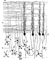
English: Section through the cerebellar cortex, parallel to the long axis of a folium.
Date image created 2009-12-15; original book published 1913
Source Figure 514 (p. 579), Cunningham's Textbook of anatomy, by Daniel John Cunningham, published in 1913 by William Wood
Author user:Looie496 created file, original artist unknown

Licensing

Public domain
Public domain
This media file is in the public domain in the United States. This applies to U.S. works where the copyright has expired, often because its first publication occurred prior to January 1, 1929, and if not then due to lack of notice or renewal. See this page for further explanation.

United States
United States
This image might not be in the public domain outside of the United States; this especially applies in the countries and areas that do not apply the rule of the shorter term for US works, such as Canada, Mainland China (not Hong Kong or Macao), Germany, Mexico, and Switzerland. The creator and year of publication are essential information and must be provided. See Wikipedia:Public domain and Wikipedia:Copyrights for more details.

Captions

Section through the cerebellar cortex, parallel to the long axis of a folium. Granule cells (GR, bottom), parallel fibers (horizontal lines, top), and Purkinje cells (P, middle) with flattened dendritic trees.

Items portrayed in this file

depicts

image/png

File history

Click on a date/time to view the file as it appeared at that time.

Date/TimeThumbnailDimensionsUserComment
current17:56, 15 December 2009Thumbnail for version as of 17:56, 15 December 2009389 × 465 (64 KB)Looie496{{Information |Description={{en|1=Section through the cerebellar cortex, parallel to the long axis of a folium. This image is figure 514 (p. 579) of Cunningham's Textbook of anatomy, by Daniel John Cunningham, published in 1913 by William Wood.}} |Source

The following page uses this file:

Global file usage

The following other wikis use this file: