File:132 (CardioNetworks ECGpedia).jpg

Original file (2,987 × 1,440 pixels, file size: 4.9 MB, MIME type: image/jpeg)
| This is a file from the Wikimedia Commons. The description on its description page there is shown below.
Commons is a freely licensed media file repository. You can help. |
.jpg)
Summary
| Description |
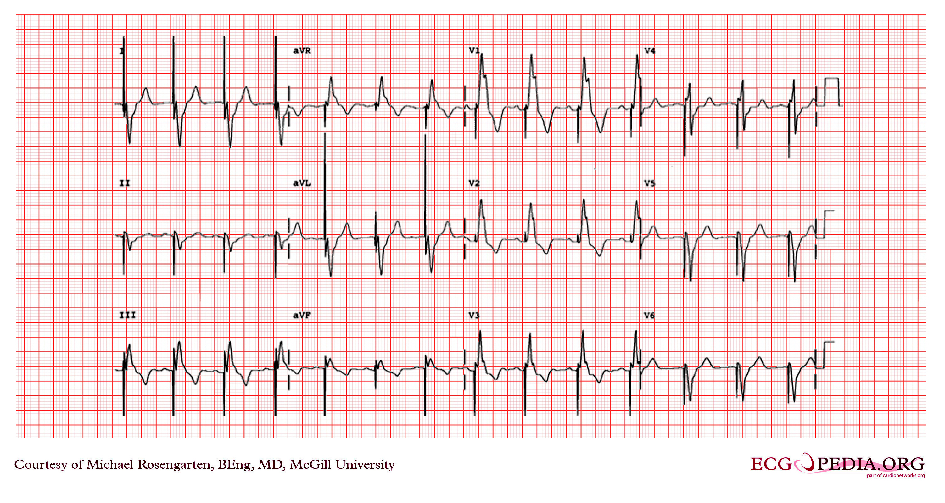
This is an electrocardiogram recorded from a patient in his 80's after implantation of a pacemaker. This recording was made as part of a routine pacemakers follow-up The pacing system was working well . This is an interesting electrocardiogram which illustrates an important point regarding pacemaker lead implantation. It is clear that this patient's pacemaker is sensing his P waves and following with pacing of his ventricle. What is curious about this recording, is the right bundle branch block configuration of the QRS and the extreme rightward axis seen in lead one. It is for this reason, a routine electrocardiogram should be done after every pacemaker implantation that involves implanting a ventricular lead. In actual fact this patient's ventricular lead was pacing the left ventricle. This lead was probably in a branch of the coronary vein . The path of the lead can be traced by the CT video which was taken after this electrocardiogram was recorded. The patient had a pacemaker system that was working well, in spite of the fact that the pacemaker was pacing the left ventricle. In fact, new pacing techniques are pacing the left ventricle as well as the right ventricle to maintain cardiac synchronization. In this case though , the ventricular lead was removed, without complication, because of an associated infection of the pacemaker pocket. A new pacemaker was implanted from the right side with the new lead placed in the right of ventricle. This resulted in a markedly different electrocardiogram. |
|
| Date | ||
| Source | EKG World Encyclopedia http://cme.med.mcgill.ca/php/index.php , courtesy of Michael Rosengarten BEng, MD.McGill | |
| Author | Michael Rosengarten BEng, MD.McGill | |
| Permission (Reusing this file) |
|
|
| CardioNetworks Source | 132.jpg |
ECGpedia Original upload log
| Date/Time | Dimensions | User | Comment |
|---|---|---|---|
| 11:09, 21 February 2012 | 3,004 × 1,599 (4.45 MB) | DarrelC (Talk • Contrib) |
File history
Click on a date/time to view the file as it appeared at that time.
| Date/Time | Thumbnail | Dimensions | User | Comment | |
|---|---|---|---|---|---|
| current | 21:59, 8 December 2013 | | 2,987 × 1,440 (4.9 MB) | Cropbot | upload cropped version, operated by User:Andy king50. Summary: cropped caption/Watermark |
| 19:41, 31 January 2013 |  | 3,004 × 1,599 (4.25 MB) | Smallbot | Commons:Batch uploading/ECGPedia: (Commons:Bots/Requests/Smallbot_8) Uploading ECGpediaCommons:Batch_uploading/AELG: Uploading photos |
File usage
The following page uses this file:
.jpg)